Biology · Ch 9 — Control and Co-ordination
The Eye: Structure and Vision
The Eye: Structure and Vision
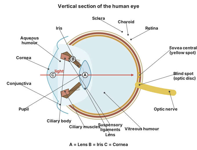
Eye : The paired eyes, the sensory organs of vision, sit in the bony orbit of the skull cushioned by fat, and each spherical eyeball is protected by the bony socket, eyebrows, eyelids, eyelashes and lacrimal (tear) glands, with six sets of muscles controlling its movement within the orbit. The eyeball's wall has three layers. The outermost, sclera (sclerotic), is a dense fibroelastic connective-tissue layer with collagen fibres that anchors the eyeball muscles; its anterior, thick, transparent, slightly bulging part is the cornea, which focuses light onto the retina, has no blood vessels of its own (it is nourished instead by aqueous humor and lacrimal secretion), and, along with the exposed sclera, is covered by the protective, lubricating conjunctiva. The middle layer, choroid (uvea), is vascular and pigmented and has three regions: the choroid proper, which lines the sclera, prevents internal light reflection through its pigment, and nourishes the retina via its blood vessels; the ciliary body, a thick, muscular ring at the choroid-iris junction whose epithelium secretes aqueous humor and whose suspensory ligaments hold and, via its muscles, adjust the lens; and the iris, formed where the choroid sharply bends inward at the sclera-cornea junction, a thin, coloured, pigment-bearing partition perforated by the pupil, whose smooth muscles regulate pupil size according to light intensity.
Lens : The lens is a transparent, elastic, biconvex structure held in place by the suspensory ligaments; together with them, it divides the eyeball's cavity into a small anterior aqueous chamber (aqueous humor) and a larger posterior vitreous chamber (vitreous humor, which also helps maintain the eyeball's shape and internal pressure). Its fine focusing adjustment for near and far objects is called accommodation.
Drawn by us to help you understand the concept clearly, and verified to make sure it's accurate. For exams, practice from your textbook's own diagram.
What this figure shows. A sectional diagram of the human eyeball labelling, front to back, the cornea and iris around the pupil, the anterior aqueous chamber filled with aqueous humor, the lens held by suspensory ligaments, the large posterior vitreous chamber filled with vitreous humor, and the three-layered wall -- outer sclera, middle vascular choroid, and inner retina -- with the fovea (region of sharpest vision), the blind spot where the optic nerve leaves, and the optic nerve itself running out the back of the eyeball.
9.19: Eye. …
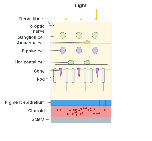
3. Retina : The innermost layer, retina, is delicate, non-vascular and light-sensitive, with a non-sensory pigmented part lining the iris/ciliary body and a sensory part lining the choroid; its inner, transparent nervous portion has three layers -- an outer photosensitive layer of rod and cone cells, a middle layer of bipolar cells, and an inner layer of ganglion cells, whose axons converge to form the optic nerve. Rods, containing the vitamin-A-derived pigment rhodopsin, handle dim-light (scotopic) vision, while three types of cones, each with its own pigment tuned to red, green or blue light, handle bright, colour (photopic) vision, with different combinations of cone stimulation producing the sensation of different colours (equal stimulation of all three gives white). Light must pass through the ganglion and bipolar layers before reaching the deeper rods and cones. The blind spot, where the optic nerve and blood vessels leave the eyeball, has no rods or cones at all; lateral to and above it, the yellow area (macula lutea) has at its centre a depression, the fovea centralis, packed with the highest density of cones and so the point of sharpest vision. To form an image, light passes through the conjunctiva, cornea, pupil and lens and is focused onto the retina; the resulting nerve impulses travel via the optic nerve to the visual area of the cerebrum, where they are analysed and the image is consciously recognised.
Drawn by us to help you understand the concept clearly, and verified to make sure it's accurate. For exams, practice from your textbook's own diagram.
What this figure shows. A magnified cross-section through the retina showing light entering from the vitreous side and having to pass through the ganglion-cell layer, then a layer of amacrine and bipolar cells, then horizontal cells, before finally reaching the outer photoreceptor layer of rod and cone cells that sits against the pigment epithelium and choroid at the very back -- so signals generated in the rods/cones travel forward again through bipolar and gangl …
Do you know?
Why do the eyes of cats and dogs glow at night? The glowing of eyes in some animals is due to a reflecting layer behind the retina, called the tapetum lucidum.
Changes in retina when light rays fall on it
- Light falls on the rod and cone cells of the retina.
- Specific wavelengths of light break up the light-sensitive pigments.
- The rod and cone cells are stimulated and a nerve impulse is generated.
- The impulse passes to the bipolar nerve cells, then to the ganglion cells. …