Q.(a) Explain the structure of immunoglobulin with suitable diagram. [figure: labelled diagram of an immunoglobulin (antibody) molecule, to be drawn by the candidate — not printed in the question paper] OR
You're viewing a preview — the full solution, concept, methods & PYQ mapping are locked.
Start your 14-day free trial to unlock the full solution →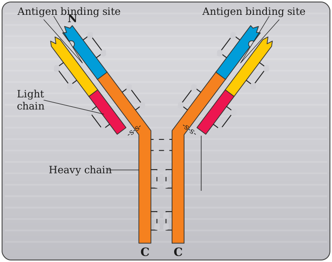
(a) A typical immunoglobulin is a Y-shaped, 4-chain (2 heavy + 2 light) protein with variable antigen-binding tips (Fab) and a constant stem (Fc). (b) Terrestrial animals show coordinated adaptations in skin, respiration, skeleton, reproduction and excretion to survive out of water.
(a) Each antibody (immunoglobulin) molecule is composed of four polypeptide chains: two identical heavy (H) chains (larger) and two identical light (L) chains (smaller), linked together by disulfide bonds into a characteristic Y-shaped structure. Along the length of each chain, the region nearer the amino-terminus/tips of the Y is called the variable (V) region -- its amino acid sequence differs from antibody to antibody, and together the variable regions of one heavy and one light chain form the antigen-binding site (paratope) that specifically recognises and binds a particular antigen's epitope, giving each antibody its unique specificity. The remainder of each chain, towards the carboxy-terminus/stem of the Y, is the constant (C) region, whose sequence is essentially the same within a given class (isotype, e.g. IgG, IgM) of antibody and determines the antibody's biological effector functions, such as its ability to bind to Fc receptors on phagocytes or to activate the complement system. Structurally, the two 'arms' of the Y, each made from one whole light chain plus the variable and first constant domain of a heavy chain, are called the Fab (fragment, antigen-binding) regions; the 'stem' of the Y, made from the remaining constant domains of the two heavy chains, is called the Fc (fragment, crystallisable) region. …
Unlock everything free for 14 days
- Full step-by-step solutions
- Concept-first explanations
- Methods, shortcuts & mistakes
- PYQ mapping + timed mock tests
Full access for 14 days. No credit card required.