Q.Draw the structure of Lymph node and label the parts. [figure: labelled diagram of a lymph node, to be drawn by the candidate — not printed in the question paper]
You're viewing a preview — the full solution, concept, methods & PYQ mapping are locked.
Start your 14-day free trial to unlock the full solution →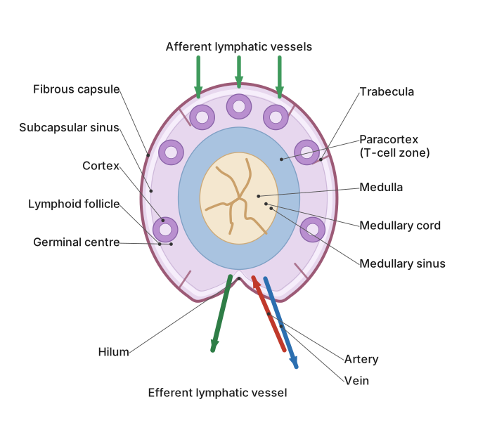
A lymph node is a bean-shaped, capsule-covered filtering organ with an outer B-cell cortex, a T-cell paracortex, and an inner medulla, receiving lymph via several afferent vessels and draining it out through one efferent vessel at the hilum.
To label the diagram correctly: draw an oval/bean-shaped structure and mark its outer covering as the fibrous capsule, from which thin connective-tissue strands called trabeculae extend inward, partitioning the interior and supporting blood vessels. Along the convex outer border, draw and label several small afferent lymphatic vessels entering the node, carrying lymph in from surrounding tissues; this lymph first passes into a thin space just beneath the capsule (the subcapsular/marginal sinus). Label the outer region of the node as the cortex, containing rounded lymphoid follicles (nodules) with pale-staining germinal centres, where B lymphocytes proliferate and differentiate; just beneath the cortex, label the paracortex (deep cortex), a T-lymphocyte-rich zone. Label the innermost region as the medulla, containing medullary cords (packed with lymphocytes and plasma cells) separated by medullary sinuses, through which the lymph, now filtered of debris/pathogens and enriched with antibodies, drains toward a single …
Unlock everything free for 14 days
- Full step-by-step solutions
- Concept-first explanations
- Methods, shortcuts & mistakes
- PYQ mapping + timed mock tests
Full access for 14 days. No credit card required.