Q.Study the given below single strand of deoxyribonucleic acid depicted in the form of a "stick" diagram with 5′ – 3′ end directionality, sugars as vertical lines and bases as single letter abbreviations and answer the questions that follow. [Stick diagram of a single polynucleotide chain showing bases A, T, G with 5′ and 3′ ends, a phosphate (P), an OH group, and covalent bonds labelled
You're viewing a preview — the full solution, concept, methods & PYQ mapping are locked.
Start your 14-day free trial to unlock the full solution →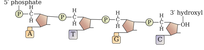
The slanting lines represent phosphodiester bonds (backbone) and N-glycosidic bonds (base-sugar linkages); the strand contains two purines (A and G); the chemical structure shows the complete sugar-phosphate backbone with attached nitrogenous bases.
The "stick" diagram is a simplified representation of a DNA polynucleotide chain, where vertical lines stand for deoxyribose sugars, single letters denote nitrogenous bases, and slanting lines indicate the covalent bonds that hold the structure together. Understanding what each bond represents is fundamental to grasping how DNA's backbone is built and how bases attach to it.
(a) Identifying the covalent bonds
Two types of covalent bonds are critical in a DNA strand, and both appear as slanting lines in stick diagrams.
Bond (a) — the slanting line connecting one sugar to the next — is a phosphodiester bond. This bond forms between the 3′ hydroxyl group of one deoxyribose sugar and the 5′ phosphate group of the next sugar, creating the sugar-phosphate backbone that runs continuously along the length of the strand. The phosphodiester linkage is what gives DNA its directional polarity: one end terminates in a free 5′ phosphate, the other in a free 3′ hydroxyl.
Bond (b) — the slanting line connecting a base to a sugar — is an N-glycosidic bond (or simply glycosidic linkage). This bond attaches the nitrogenous base to the 1′ carbon of the deoxyribose sugar. It forms between the nitrogen atom at position 9 of a purine (or position 1 of a pyrimidine) and the sugar, anchoring each base to the backbone.
The phosphodiester bond is the "spine" of DNA, while the N-glycosidic bond is the "hinge" that holds each base onto that spine.
(b) Counting the purines
Purines are the larger nitrogenous bases with a double-ring structure: adenine (A) and guanine (G). Pyrimidines — cytosine (C), thymine (T), and uracil (U, in RNA) — have a single ring.
Looking at the given strand with bases A, T, and G:
- A (adenine) is a purine.
- T (thymine) is a pyrimidine.
- G (guanine) is a purine.
There are two purines in the diagram: one adenine and one guanine.
Always remember: A and G are purines (double rings); C, T, and U are pyrimidines (single rings). This distinction is essential for understanding base pairing and Chargaff's rules.
(c) Drawing the chemical structure
A complete chemical structure of the polynucleotide chain must show:
- Deoxyribose sugars — five-membered rings (furanose form) with carbons numbered 1′ to 5′. The 2′ carbon lacks a hydroxyl group (hence "deoxy").
- Phosphate groups — linking the 3′ carbon of one sugar to the 5′ carbon of the next via ester bonds (the phosphodiester linkage).
- Nitrogenous bases — attached to the 1′ carbon of each sugar via N-glycosidic bonds. …
Unlock everything free for 14 days
- Full step-by-step solutions
- Concept-first explanations
- Methods, shortcuts & mistakes
- PYQ mapping + timed mock tests
Full access for 14 days. No credit card required.