Q.The following question is a case-based question. Read the case carefully and answer the questions that follow. Ribose and 2-deoxyribose have an important role in biology. Among the most important derivatives are those with phosphate groups attached at the 5 position. Mono-, di- and tri-phosphate forms are important, as well as 3-5 cyclic monophosphates. Purines and pyrimidines form an important class of compounds with ribose and deoxyribose. When these purine and pyrimidine derivatives are coupled to a ribose sugar, they are called nucleosides. Answer the following questions :
You're viewing a preview — the full solution, concept, methods & PYQ mapping are locked.
Start your 14-day free trial to unlock the full solution →Concept understanding — Nucleic Acid Components
Nucleic Acid Components: The Alphabet of Life
Imagine you want to write a book that contains all the instructions for building and running a living organism. You'd need an alphabet — a set of letters that can be combined in endless ways to form words, sentences, and chapters. In living cells, that alphabet is made of nucleic acids (DNA and RNA), and each "letter" is called a nucleotide.
The Big Picture: What Are Nucleic Acids?
Nucleic acids are long, chain-like molecules that store and transmit genetic information. DNA holds the master blueprint; RNA helps execute it. But both are built from the same basic building block: the nucleotide.
Think of a nucleotide as a single Lego brick. DNA and RNA are long chains of these bricks, each brick slightly different from the next.
The Three Parts of a Nucleotide
Every nucleotide has three components, like a three-part key:
- A phosphate group — a small, negatively charged group (). It acts like the "handle" that links nucleotides together.
- A sugar — either ribose (in RNA) or deoxyribose (in DNA). This is the "body" of the brick.
- A nitrogenous base — a ring-shaped molecule containing nitrogen. This is the "colored part" that carries the actual information.
The sugar and base together form a nucleoside. When you add the phosphate, you get a nucleotide.
Nucleoside = Sugar + Base
Nucleotide = Sugar + Base + Phosphate
The Two Families of Bases
The bases come in two structural types:
- Purines (double-ring structures): Adenine (A) and Guanine (G)
- Pyrimidines (single-ring structures): Cytosine (C), Thymine (T) (only in DNA), and Uracil (U) (only in RNA)
A mnemonic: Purines are Pure as All Gold (A and G). Pyrimidines are CUT (C, U, T).
DNA vs. RNA: The Key Differences
| Feature | DNA | RNA |
|---|---|---|
| Sugar | Deoxyribose (missing one oxygen) | Ribose (has that oxygen) |
| Bases | A, G, C, T | A, G, C, U |
| Structure | Double-stranded helix | Usually single-stranded |
| Function | Stores genetic information | Carries and executes instructions |
A common mistake: thinking "nucleoside" and "nucleotide" are the same. Remember: nucleotide has the phosphate; nucleoside does not. ATP (adenosine triphosphate) is a nucleotide — it's the energy currency of the cell.
Why This Matters
The sequence of bases along a DNA strand spells out the genetic code. A change in even one base (a mutation) can alter a protein, sometimes with dramatic consequences — like sickle cell anemia, where a single base change in the hemoglobin gene causes red blood cells to deform. …
Why this formula?
Nucleic Acid Components: Understanding the "Why" Behind the Key Relationships
Let’s start with the big picture: Nucleic acids (DNA and RNA) are polymers made of nucleotides. Each nucleotide has three parts: a nitrogenous base, a pentose sugar, and a phosphate group. The key formulas and relationships in this topic arise from how these parts are linked and how they behave chemically.
1. The Basic Composition Formula
What it says:
A nucleotide = Base + Sugar + Phosphate
Why this holds:
- Chemical necessity: The sugar (ribose in RNA, deoxyribose in DNA) has a 5-carbon ring. The base attaches to the 1' carbon (via a glycosidic bond), and the phosphate attaches to the 5' carbon (via an ester bond).
- Directionality: This creates a 5' → 3' linkage in the polymer. The phosphate of one nucleotide bonds to the 3' carbon of the next sugar.
- Reasoning: Without the phosphate, you have a nucleoside (base + sugar). Adding the phosphate makes it a nucleotide — the monomer that can polymerize.
Key takeaway: The formula isn’t arbitrary — it reflects the specific carbon positions on the sugar that allow for stable, directional chain formation.
2. Chargaff’s Rules (for DNA)
What it says:
In double-stranded DNA:
Why this holds:
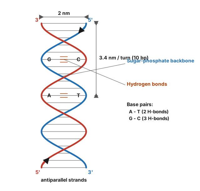
- Base pairing: Adenine (A) forms two hydrogen bonds with Thymine (T). Guanine (G) forms three hydrogen bonds with Cytosine (C).
- Structural constraint: The DNA double helix has a constant width (20 Å). A purine (A or G) always pairs with a pyrimidine (T or C) — otherwise the helix would bulge or narrow.
- Derivation: If every A on one strand must pair with a T on the opposite strand, then the number of A equals number of T in the whole molecule. Same for G and C.
- Consequence: The sum of purines equals sum of pyrimidines ().
Why it’s not just a rule: It’s a geometric and energetic necessity — hydrogen bonding and helix stability force this equality.
3. The Phosphodiester Bond Energy Formula
What it says:
Formation of a phosphodiester bond requires ~30 kJ/mol of energy (from ATP).
Why this holds:
- Mechanism: The 3' hydroxyl of one nucleotide attacks the α-phosphate of a nucleotide triphosphate (e.g., ATP). This releases pyrophosphate (PPi).
- Energy source: The hydrolysis of PPi to two inorganic phosphates () is highly exergonic (ΔG ≈ -30 kJ/mol). This drives the reaction forward.
- Reasoning: The bond itself is a covalent ester linkage — strong but not spontaneously formed. The energy comes from breaking a high-energy phosphate bond in the triphosphate.
Key insight: The formula isn’t about the bond’s strength — it’s about the thermodynamic cost of making it in a cell.
4. The Melting Temperature () Formula
What it says:
(in °C) ≈ for short oligonucleotides.
Why this holds:
- Hydrogen bonds: G-C pairs have 3 H-bonds, A-T pairs have 2 H-bonds. More H-bonds = more energy needed to separate strands.
- Stacking interactions: G-C base pairs also have stronger π-stacking (aromatic ring overlap) than A-T.
- Derivation: The formula is empirical — it comes from measuring for many sequences. The coefficients (4 and 2) reflect the relative stability contributed by each base pair.
- Limitation: For long DNA, this simple formula fails because nearest-neighbor interactions matter more.
Why it works: It’s a linear approximation of the free energy needed to break all base pairs, weighted by H-bond count.
5. The Central Dogma (Information Flow)
What it says:
DNA → RNA → Protein
Why this holds: …
(a) DNA hydrolysis → 2‑deoxyribose + phosphoric acid + bases A, G, C, T; DNA differs from RNA in having deoxyribose (not ribose) and thymine (not uracil), and is double‑stranded. (b) Nucleoside = base + sugar, nucleotide = base + sugar + phosphate. (c)(i) Nucleic acids store genetic information and direct protein synthesis. OR (c)(ii) The joining linkage is a 3′–5′ phosphodiester bond; the RNA‑only base is uracil.
Every nucleic acid is a polymer of nucleotides, each built from a nitrogenous base, a pentose sugar and a phosphate group. Hydrolysis simply reverses that assembly.
Products of DNA hydrolysis. Water breaks two kinds of bonds — the phosphodiester bonds between nucleotides and the glycosidic bonds between sugar and base. Complete hydrolysis therefore liberates:
- 2‑deoxyribose (a 5‑carbon sugar lacking the –OH at C‑2′),
- phosphoric acid,
- the four bases adenine, guanine, cytosine and thymine.
List thymine, not uracil, for DNA. Uracil belongs to RNA.
How DNA differs from RNA structurally.
| Feature | DNA | RNA |
|---|---|---|
| Sugar | 2‑deoxyribose (no 2′‑OH) | ribose (2′‑OH present) |
| Bases | A, G, C, T | A, G, C, U |
| Strands | double helix (antiparallel) | usually single |
The 2′‑OH of ribose makes RNA more reactive and less stable, which is why DNA is the long‑term store of information.
Concept understanding — Biochemical Bonds
Biochemical Bonds: The Glue That Holds Life Together
Imagine you're building with LEGO bricks. Some bricks click together tightly and never come apart unless you really yank them. Others snap together lightly and can be pulled apart with a gentle tug. Some bricks don't even click — they just stick because of static cling or magnetism.
Biochemical bonds are exactly like that. They are the forces that hold atoms together inside the molecules of your body — your DNA, proteins, fats, and carbohydrates. Without these bonds, you'd literally fall apart into a pile of individual atoms.
The Core Idea
Atoms bond because being bonded is more stable (lower energy) than being alone. Think of it like this: a single atom is like a person standing alone in a cold room. Bonding is like huddling together for warmth — you lose some freedom of movement, but you gain stability.
In biochemistry, we care about four main types of bonds. They differ in strength, how they form, and what they do in living systems.
1. Covalent Bonds — The Strong, Permanent LEGO Clicks
This is the strongest bond in biology. Two atoms share electrons — like two people holding the same umbrella. Each atom contributes one or more electrons, and they both "own" the pair.
Key properties:
- Very strong (100–400 kJ/mol)
- Forms the backbone of all biomolecules
- Takes a lot of energy (or enzymes) to break
Where you find it:
- The carbon-carbon bonds in your DNA's sugar-phosphate backbone
- The peptide bonds linking amino acids into proteins
- The bonds within a glucose molecule
A single covalent bond shares 2 electrons. A double bond shares 4. Triple bonds are rare in biology but exist (e.g., in cyanide).
2. Ionic Bonds — The Static Cling of Opposites
Some atoms steal electrons from others. When that happens, one atom becomes positively charged (lost an electron) and the other becomes negatively charged (gained one). Opposite charges attract — that's an ionic bond.
Key properties:
- Moderate strength (5–100 kJ/mol in dry conditions)
- Very weak in water (because water molecules get in between)
- Easily broken by changes in pH or salt concentration
Where you find it:
- In salt bridges that help proteins fold into their correct shape
- Between the phosphate groups of DNA and positively charged proteins (histones)
Ionic bonds are often called "bonds" but in water they behave more like attractions. Don't confuse them with covalent bonds — they're much weaker in biological fluids.
3. Hydrogen Bonds — The Gentle, Reversible Magnets
This is the most important weak bond in biology. A hydrogen atom that's already covalently bonded to an electronegative atom (like oxygen or nitrogen) gets a slight positive charge. It then gets attracted to another electronegative atom nearby.
Think of it like a weak magnet — it holds things together but can be easily undone.
Key properties:
- Weak individually (5–30 kJ/mol)
- But many together can be very strong
- Easily broken by heat or changes in pH
- Directional — they only work when atoms are properly aligned
Where you find it:
- Between the two strands of DNA (this is what holds the double helix together)
- In protein folding (between amino acids in the backbone)
- Between water molecules (giving water its unique properties)
Hydrogen bonds are the reason DNA can unzip for replication. If DNA used covalent bonds between strands, it would be impossible to separate without destroying the molecule.
4. Van der Waals Interactions — The Fleeting, Accidental Touches
Even neutral atoms have temporary, uneven distributions of electrons. These create tiny, momentary charges that attract nearby atoms. It's like two people accidentally brushing shoulders in a crowd — brief, weak, but real.
Key properties:
- Extremely weak (0.5–5 kJ/mol per interaction)
- Only work when atoms are very close (within 0.3–0.4 nm)
- Add up significantly when many atoms are packed together
Where you find it:
- In the hydrophobic core of proteins (where oily amino acids pack tightly)
- Between lipid tails in cell membranes
- In enzyme-substrate binding (helps "grip" the substrate)
Putting It All Together: A Biological Example
Consider a protein in your body. It's a long chain of amino acids held together by covalent peptide bonds. That chain then folds into a specific shape. The folding is guided by:
- Hydrogen bonds between backbone atoms (forming alpha helices and beta sheets)
- Ionic bonds between charged side chains …
Why this formula?
Biochemical Bonds: Why the Key Formulas Hold
Biochemical bonds are the forces that hold atoms together in biomolecules. The key formulas come from electrostatics and quantum mechanics — not from biology itself. Let's break down the why behind the most important ones.
1. Ionic Bond Energy: Coulomb's Law
Formula:
Why it holds:
- Opposite charges attract — this is a fundamental law of physics (Coulomb's law).
- In a biochemical context, consider a sodium ion () and a chloride ion (). The energy released when they come together is directly proportional to the product of their charges () and inversely proportional to the distance () between them.
- The constant accounts for the medium (water vs. vacuum). In water, the effective force is weaker because water molecules partially shield the charges — this is why ionic bonds in biology are often weaker in aqueous environments.
Key insight: The formula is not arbitrary — it's derived from the inverse-square law of electrostatics, integrated over the distance the charges move toward each other.
2. Covalent Bond Energy: The Morse Potential (Approximation)
Formula (simplified):
Why it holds:
-
Covalent bonds arise from shared electrons between atoms. The energy is not a simple inverse-square law because electrons are delocalized.
-
The Morse potential is an empirical formula that captures two key observations:
- At equilibrium distance (): Energy is minimum ( in this form).
- If atoms are pulled apart (): Energy approaches (the bond dissociation energy).
- If atoms are pushed too close (): Energy skyrockets due to Pauli repulsion (electrons can't occupy the same space).
-
The exponential term models the rapid drop in attractive force as distance increases — this comes from quantum mechanical overlap of electron clouds.
Key insight: The formula is a curve fit to quantum mechanical calculations, not a first-principles derivation. But it works because it respects the physics: attraction at long range, repulsion at short range, and a stable minimum.
3. Hydrogen Bond Energy: Dipole-Dipole Interaction
Formula (approximate):
Why it holds:
- A hydrogen bond (e.g., between water molecules) is not a true bond — it's a strong dipole-dipole interaction.
- The dipole moment () arises because oxygen is more electronegative than hydrogen, creating partial charges ( and ).
- The energy depends on:
- Strength of dipoles ()
- Distance () — falls off as , much faster than ionic bonds ()
- Orientation () — strongest when dipoles are aligned head-to-tail
Key insight: The dependence comes from the derivative of the dipole field. Unlike point charges, dipoles have a field that decays faster — this is why hydrogen bonds are directional and weaker than covalent bonds.
4. Van der Waals Interaction: Lennard-Jones Potential
Formula:
Why it holds:
- Van der Waals forces arise from temporary fluctuations in electron distribution — even nonpolar molecules have instantaneous dipoles. …
(a) DNA hydrolysis → 2‑deoxyribose + phosphoric acid + bases A, G, C, T; DNA differs from RNA in having deoxyribose (not ribose) and thymine (not uracil), and is double‑stranded. (b) Nucleoside = base + sugar, nucleotide = base + sugar + phosphate. (c)(i) Nucleic acids store genetic information and direct protein synthesis. OR (c)(ii) The joining linkage is a 3′–5′ phosphodiester bond; the RNA‑only base is uracil.
Every nucleic acid is a polymer of nucleotides, each built from a nitrogenous base, a pentose sugar and a phosphate group. Hydrolysis simply reverses that assembly.
(b) Nucleoside vs nucleotide
- Nucleoside = nitrogenous base + pentose sugar only (e.g. adenosine, deoxyadenosine). The base is attached to C‑1′ of the sugar by a β‑glycosidic bond.
- Nucleotide = nucleoside + one or more phosphate groups esterified at C‑5′ (e.g. AMP, dATP).
"‑oside" = sugar + base only; "‑otide" = oside + phosphate.
Concept understanding — this question
Explanation coming soon.
(a) DNA hydrolysis → 2‑deoxyribose + phosphoric acid + bases A, G, C, T; DNA differs from RNA in having deoxyribose (not ribose) and thymine (not uracil), and is double‑stranded. (b) Nucleoside = base + sugar, nucleotide = base + sugar + phosphate. (c)(i) Nucleic acids store genetic information and direct protein synthesis. OR (c)(ii) The joining linkage is a 3′–5′ phosphodiester bond; the RNA‑only base is uracil.
Every nucleic acid is a polymer of nucleotides, each built from a nitrogenous base, a pentose sugar and a phosphate group. Hydrolysis simply reverses that assembly.
(c)(i) Two functions of nucleic acids
- Storage and transmission of genetic information — the base sequence of DNA encodes genes passed from parent to offspring. …
Unlock everything free for 14 days
- Full step-by-step solutions
- Concept-first explanations
- Methods, shortcuts & mistakes
- PYQ mapping + timed mock tests
Full access for 14 days. No credit card required.