Biology · Ch 5 — Origin and Evolution of Life
B. Morphology
B. Morphology
B. Morphology : Morphology deals with the study of external structures, while anatomy deals with the study of internal structures. From the comparative study of morphology and anatomy we can understand the evolutionary aspects in the form of homologous, analogous and vestigial organs.
a. Homologous organs : Homologous organs are those organs which are structurally similar but perform different functions. For example:
- The forelimbs of vertebrates such as the lizard, bird, bat, horse, whale and man: all of them have a humerus, radius-ulna, carpals, metacarpals and phalanges in their forelimbs. The forelimbs of these vertebrates are structurally similar but perform different functions.
- The vertebrate heart and brain.
- In plants, the thorns of Bougainvillea and the tendrils of Cucurbita represent homology; the thorns of Bael and the tendrils of the passion flower are homologous.
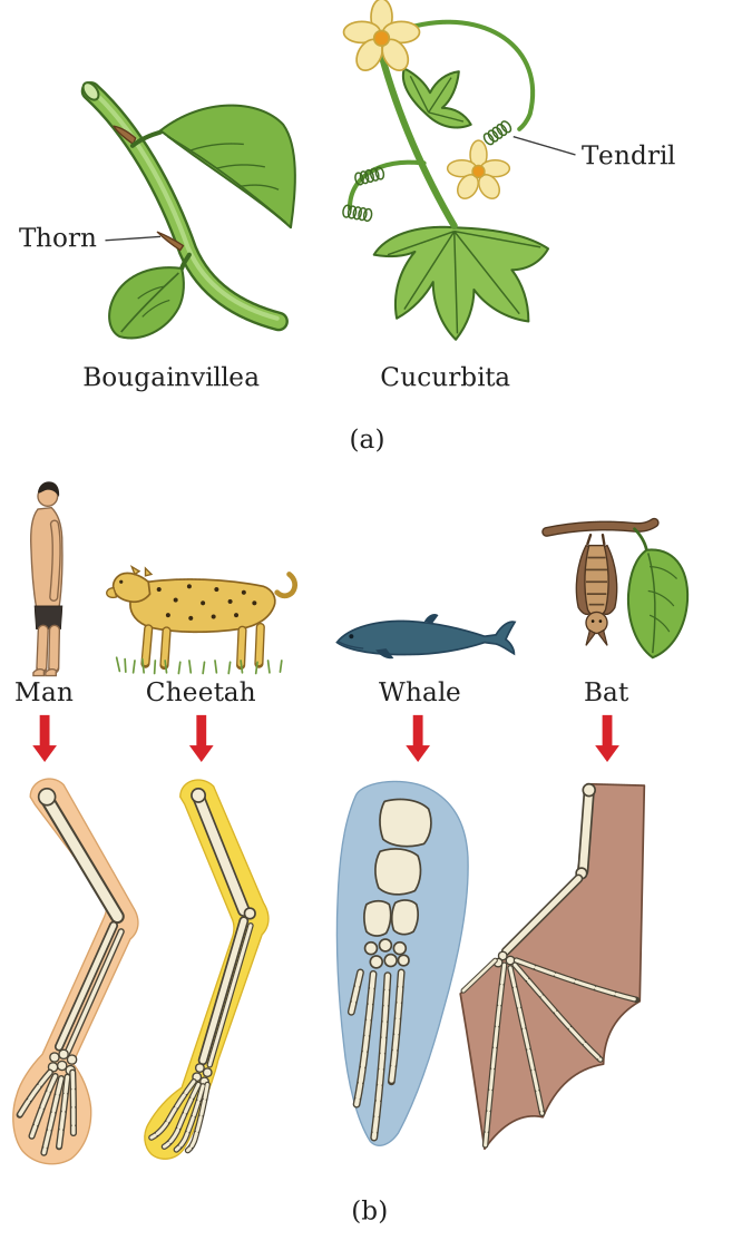
Drawn by us to help you understand the concept clearly, and verified to make sure it's accurate. For exams, practice from your textbook's own diagram.
What this figure shows. A comparison of the forelimb skeletons of a lizard, bird, bat, horse, whale and human laid out side by side, each labelled to show the same underlying bone set — humerus, radius, ulna, carpals, metacarpals and phalanges — even though the overall shape of each limb has been reshaped very differently for walking, flying, swimming or grasping; alongside it, a plant panel comparing the thorn of Bougainvillea with the tendr …
The structural similarities between homologous organs indicate that they have a common ancestry. Differences in homologous organs are examples of divergent evolution or adaptive radiation.
b. Analogous organs : Analogous organs are those which are structurally dissimilar but functionally similar. They have an external, superficial similarity due to similar functions but are different anatomically, e.g. the wings of a butterfly (insect) and of a bird look superficially alike but are not anatomically similar structures, though they perform similar functions. Other examples of analogous organs:
- The eye of the octopus (mollusca) and of mammals; they differ in the position of the retina, the structure of the lens and the origin of the different eye parts.
- The flippers of penguins (birds) and dolphins (mammals).
- Sweet potato (a root modification) and potato (a stem modification), which store food in the form of starch.
- The terminal leaflets of pea and the apical bud of Vitis, which are modified into tendrils.
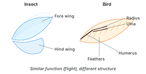
Drawn by us to help you understand the concept clearly, and verified to make sure it's accurate. For exams, practice from your textbook's own diagram.
What this figure shows. A side-by-side comparison of an insect's wing (a thin membranous fore-wing and hind-wing with no internal bones) and a bird's wing (a feathered structure built around a humerus, radius and ulna), visually emphasising that although both structures serve the same function of flight and look superficially similar in outline, their internal anatomy is completely different, showing they evolved indepen …
Analogous organs lead to convergent evolution, i.e. different organisms show the same superficial structural similarities due to similar functions or habitat. These organs do not help to trace common ancestry; thus analogous organs do not have a significant role in evolution.
c. Vestigial organs (Rudimentary organs) : Vestigial organs are imperfectly developed, non-functional, degenerate structures which were functional in some related or other animals or in ancestors. They are no longer required by the organism but indicate its relationship with those organisms in which these organs are fully developed. Examples: human beings show vestigial organs like -
- Presence of a vestigial nictitating membrane (the plica semilunaris).
- Presence of wisdom teeth (third molars).
- Coccyx (tail bone): greatly reduced in man, since the tail is of no use because of the erect posture. …
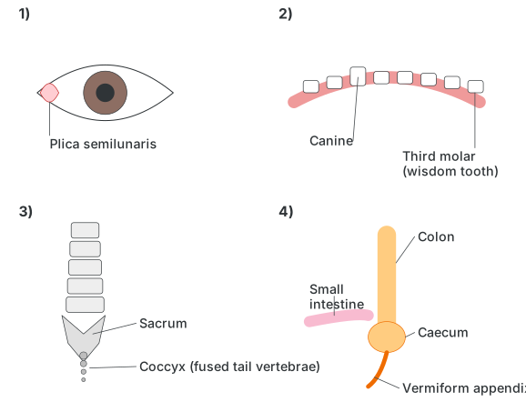
Drawn by us to help you understand the concept clearly, and verified to make sure it's accurate. For exams, practice from your textbook's own diagram.
What this figure shows. A labelled diagram of the human body highlighting four vestigial structures in their approximate anatomical locations: the plica semilunaris (a small fold at the inner corner of the eye, the vestige of a nictitating membrane), the canine and third molar/wisdom tooth in the jaw, the sacrum with the small fused coccyx (tail vertebrae) at its base, and the caecum with its narrow vermiform appendix branching …