Q.Draw and label the parts of Cycas Rachis T.S.
You're viewing a preview — the full solution, concept, methods & PYQ mapping are locked.
Start your 14-day free trial to unlock the full solution →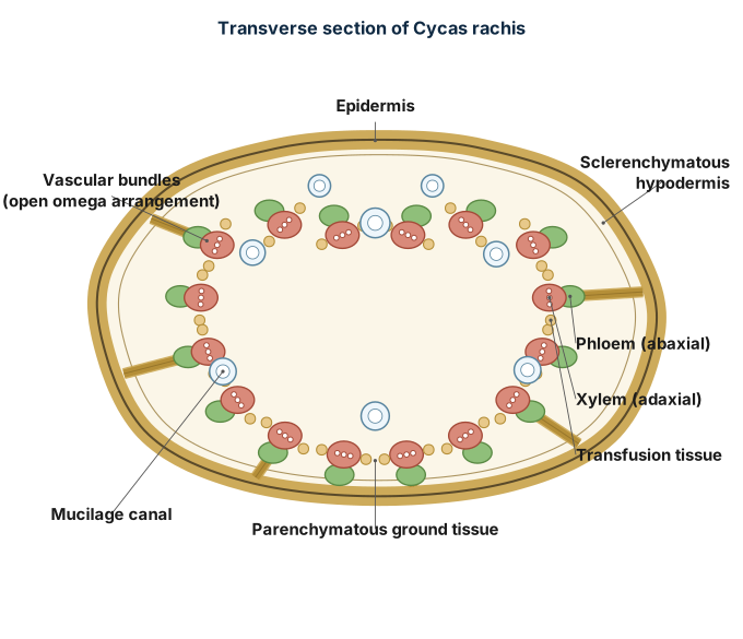
This question asks for a labelled drawing, which cannot be rendered as an image here; instead, the anatomy of a Cycas rachis in transverse section is described part-by-part below so the same labelled structure can be reproduced as a diagram.
A transverse section (T.S.) of the rachis (the main axis of a Cycas pinnate leaf) shows, from the outside inward, the following regions — draw and label each of these in your diagram:
-
Epidermis — the outermost single layer of compactly arranged, cutinized cells, protecting the rachis.
-
Hypodermis — a multi-layered zone of sclerenchymatous cells just beneath the epidermis, providing mechanical strength; note that girder-like extensions of hypodermal sclerenchyma often occur above and below the vascular bundles.
-
Ground tissue (cortex) — a wide zone of thin-walled parenchyma cells occupying the bulk of the rachis, in which scattered mucilage (resin) canals are embedded — a distinctive feature of Cycas.
-
Vascular bundles — arranged in a characteristic horseshoe or omega (Ω)-shaped ring within the ground tissue (unlike the scattered arrangement in monocot stems or the single ring in typical dicot stems). Each individual vascular bundle is collateral and open, and consists of:
- Xylem — located toward the inner/adaxial side, made up mainly of tracheids, with associated transfusion tissue (short tracheid-like cells that help lateral conduction of water, characteristic of gymnosperms). …
Unlock everything free for 14 days
- Full step-by-step solutions
- Concept-first explanations
- Methods, shortcuts & mistakes
- PYQ mapping + timed mock tests
Full access for 14 days. No credit card required.