Question 43 of 56
Q.Draw a neat labelled diagram of L.S. of the human eye.
Tamil Nadu DgeTamil Nadu HSC First Year (DGE) Board 2023Subjective· 3mImportance★★★★★
77% · 43/56 Questions
You're viewing a preview — the full solution, concept, methods & PYQ mapping are locked.
Start your 14-day free trial to unlock the full solution →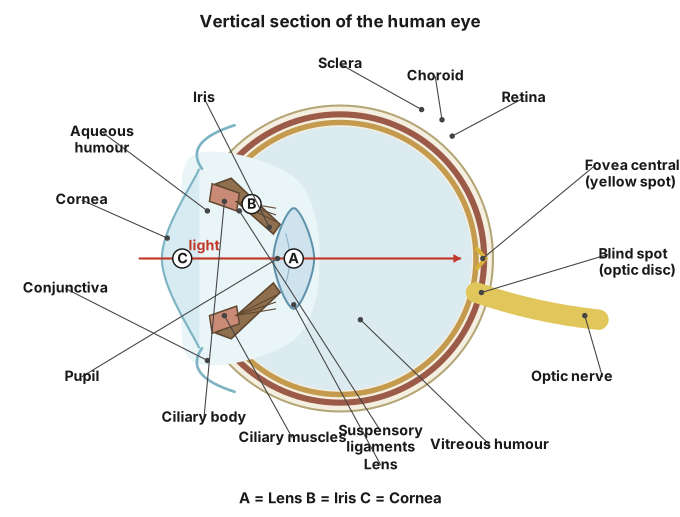
A labelled L.S. of the human eye shows three coats (sclera/cornea, choroid/ciliary body/iris, retina), the lens, its two fluid-filled chambers, and the optic nerve exit point — described here in place of a drawn figure.
Since this format cannot render an actual drawing, the required labelled structure is described part by part, in the order it would be drawn and labelled in a longitudinal (L.S.) diagram of the eyeball:
Outer (fibrous) coat:
- Sclera — the tough, opaque white outer layer covering most of the eyeball, giving it shape and protection.
- Cornea — the transparent, curved front continuation of the sclera, through which light first enters and is partly focused.
Middle (vascular) coat:
- Choroid — a dark, pigmented, richly vascular layer that nourishes the eye and absorbs stray light.
- Ciliary body — a thickened anterior extension of the choroid, holding the lens in place via suspensory ligaments and controlling its shape (accommodation).
- Iris — the pigmented, muscular diaphragm in front of the lens, with a central opening.
- Pupil — the opening in the centre of the iris that regulates the amount of light entering the eye.
Lens:
- A transparent, biconvex structure held behind the iris by suspensory ligaments from the ciliary body, focusing light onto the retina.
Chambers:
- Aqueous humour — watery fluid filling the space between cornea and lens (anterior chamber).
- Vitreous humour — thick, jelly-like fluid filling the space between lens and retina (posterior chamber), maintaining the eyeball's shape.
Inner (nervous) coat: …
Unlock everything free for 14 days
- Full step-by-step solutions
- Concept-first explanations
- Methods, shortcuts & mistakes
- PYQ mapping + timed mock tests
Full access for 14 days. No credit card required.