Q.(a) Explain the structure of neuron with a labelled diagram. OR
You're viewing a preview — the full solution, concept, methods & PYQ mapping are locked.
Start your 14-day free trial to unlock the full solution →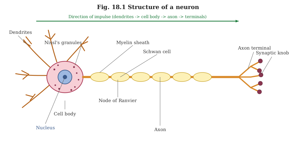
A neuron is built from a cell body (cyton), dendrites that receive signals, and an axon (often myelinated) that carries signals away to synaptic terminals — described here in place of a drawn, labelled figure.
(a) Structure of a neuron:
Since this format cannot render an actual drawing, the structure is described part by part, matching what a correctly labelled diagram would show:
-
Cyton (cell body/soma): the main body of the neuron, containing the nucleus and cytoplasm. The cytoplasm contains characteristic granular structures called Nissl granules (rough endoplasmic reticulum with ribosomes), which are the site of protein synthesis.
-
Dendrites: short, thin, branched processes that arise from the cyton. They receive nerve impulses from other neurons (or receptor cells) and conduct these impulses toward the cell body.
-
Axon: a single, typically much longer process arising from the cyton (at a region called the axon hillock), which conducts the nerve impulse away from the cell body toward another neuron, muscle or gland.
- Many axons are covered by a fatty, insulating myelin sheath, formed by wrapping Schwann cells (in the peripheral nervous system) around the axon.
- The myelin sheath is interrupted at regular intervals by small gaps called Nodes of Ranvier, where the axon membrane is exposed. These nodes allow the nerve impulse to 'jump' from node to node (saltatory conduction), greatly increasing the speed of conduction compared to an unmyelinated axon. …
Unlock everything free for 14 days
- Full step-by-step solutions
- Concept-first explanations
- Methods, shortcuts & mistakes
- PYQ mapping + timed mock tests
Full access for 14 days. No credit card required.