Q.Given below are the diagrammatic representations of the replicating fork of DNA in E. coli. Study the diagrams and answer the questions that follow. [Diagram (i): fork labelled 3' and 5' at top, 5' and 3' at bottom; Diagram (ii): fork labelled 5' and 3' at top, 3' and 5' at bottom; Diagram (iii): fork labelled 3' and 5' at top, 5' and 3' at bottom]
You're viewing a preview — the full solution, concept, methods & PYQ mapping are locked.
Start your 14-day free trial to unlock the full solution →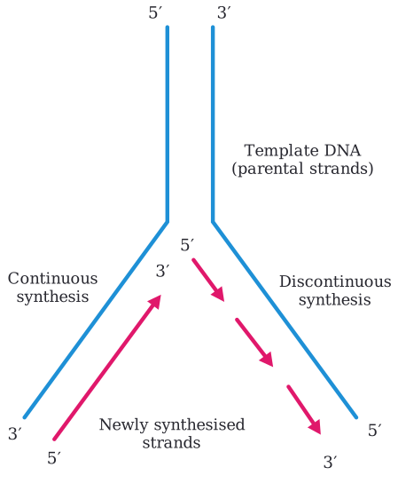
Of diagrams (i) and (iii) -- which the source figure labels identically -- whichever one shows continuous 5' to 3' synthesis on the leading strand and discontinuous 5' to 3' synthesis (Okazaki fragments) on the lagging strand is correct; diagram (ii) is definitely wrong (reversed template polarity). The enzyme DNA ligase joins the newly synthesized DNA fragments.
DNA replication is a fundamental process where a cell makes an exact copy of its DNA. To understand the diagrams, we need to recall the core principles governing this process. The DNA double helix consists of two strands that are anti-parallel, meaning one runs 5' to 3' and the other runs 3' to 5'. During replication, these strands separate, and each serves as a template for a new complementary strand.
The crucial point is that DNA polymerase, the enzyme responsible for synthesizing new DNA, can only add nucleotides to the 3'-hydroxyl end of a growing strand. This means new DNA is always synthesized in the 5' to 3' direction.
Because the two template strands are anti-parallel, and synthesis must always be 5' to 3', DNA replication proceeds differently on each template:
- Leading strand: On the template strand that runs 3' to 5' (relative to the replication fork), the new strand can be synthesized continuously in the 5' to 3' direction, moving towards the replication fork.
- Lagging strand: On the template strand that runs 5' to 3' (relative to the replication fork), the new strand must be synthesized discontinuously in short segments called Okazaki fragments. Each fragment is synthesized 5' to 3', but in a direction away from the replication fork. These fragments are later joined together.
Let's analyze the given diagrams based on these principles.
Part (a): Identifying the Correct Diagram
-
Analyze Diagram (ii):
- The top template strand runs 5' to 3' (left to right); the bottom template runs 3' to 5' (left to right) -- the reverse polarity from (i)/(iii).
- Whichever strand-synthesis pattern is correct for (i)/(iii), diagram (ii)'s reversed template labeling makes it the odd one out and definitely wrong.
-
Diagrams (i) and (iii) -- an honest note on this figure's transcription:
Both diagrams are labeled identically in the source transcription available here (top strand 3' to 5', bottom strand 5' to 3'), with no further detail captured on how the newly synthesized strand is actually drawn in each (continuous line vs. separate fragments). That distinguishing detail exists on the real printed exam figure but was not captured in this transcription, so this answer cannot honestly assert which specific one of (i)/(iii) is correct.
Watch outA common mistake is to assume the lagging strand is synthesized 3' to 5' because its template is 5' to 3'. Remember, each individual Okazaki fragment is synthesized 5' to 3'. The overall direction of growth for the lagging strand away from the fork is a consequence of this 5' to 3' synthesis on a 5' to 3' template. …
Unlock everything free for 14 days
- Full step-by-step solutions
- Concept-first explanations
- Methods, shortcuts & mistakes
- PYQ mapping + timed mock tests
Full access for 14 days. No credit card required.