Q.Describe the structure of an ideal antibody molecule with diagram.
You're viewing a preview — the full solution, concept, methods & PYQ mapping are locked.
Start your 14-day free trial to unlock the full solution →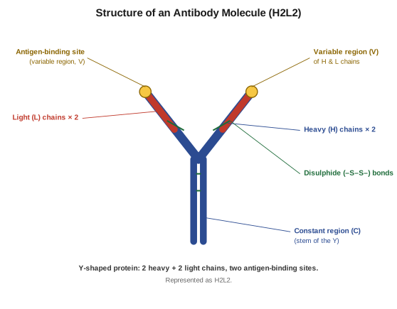
An antibody is a Y-shaped H2L2 protein: two heavy + two light chains joined by disulphide bonds, with two variable-region antigen-binding sites at the tips of the arms and a constant-region stem.
Structure of an ideal antibody molecule:
-
Four polypeptide chains: Every antibody is made of four chains — two identical heavy (H) chains (longer) and two identical light (L) chains (shorter). This is written as H2L2.
-
Shape: The chains are arranged in a Y-shape. The two light chains lie along the two arms; the two heavy chains run through the arms and the stem.
-
Disulphide bonds: The chains are linked to one another by disulphide (–S–S–) bonds.
-
Variable (V) and constant (C) regions: The tip of each arm has a variable region (its amino-acid sequence differs from antibody to antibody); together the variable regions of an H and an L chain form an antigen-binding site. So each antibody has two antigen-binding sites. The rest of the molecule is the constant region.
…
Unlock everything free for 14 days
- Full step-by-step solutions
- Concept-first explanations
- Methods, shortcuts & mistakes
- PYQ mapping + timed mock tests
Full access for 14 days. No credit card required.