Q.(a) Draw a sectional view of human ovary. Label the following parts :
🔒You're viewing a preview — the full solution, concept, methods & PYQ mapping are locked.
🔒 Start your 14-day free trial to unlock the full solution →Part (a)Concept understanding — Reproductive System Functions
Reproductive System Functions – A First Look
Think about what every living thing does, without exception: it reproduces. A mango tree grows from a seed, flowers, and produces more mangoes. A cat gives birth to kittens. You yourself are here because two people, your parents, reproduced. Reproduction is the biological process by which organisms create new individuals of their own kind — it is the reason life continues across generations.
For a commerce or humanities student, the reproductive system is not about complicated diagrams or medical terms. It is about understanding the purpose and basic working of the system that ensures the survival of the human species. The NCERT textbook presents this as a fundamental life process, just like nutrition, respiration, or excretion.
What Does the Reproductive System Do?
The primary function of the reproductive system is to produce offspring. But that simple statement hides a lot of careful design. The system must:
- Produce gametes — the male sperm and the female egg (ovum).
- Transport these gametes so they can meet and fertilise.
- Provide a safe environment for the development of the fertilised egg into a baby.
- Nourish the newborn after birth (through milk production in females).
In humans, reproduction is sexual — it requires two parents, one male and one female. This is different from asexual reproduction (like a bacterium splitting into two), and it introduces genetic variation, which is why brothers and sisters are not identical copies of each other.
The reproductive system is the only organ system that is not essential for the survival of the individual — you can live without reproducing. But it is essential for the survival of the species. That is why it is considered a life process in biology.
The Male Reproductive System – Key Functions
The male system is designed to produce and deliver sperm. The NCERT textbook highlights these main parts and their roles:
- Testes (two oval organs): Produce sperm and the male hormone testosterone. Testosterone is responsible for changes during puberty — deeper voice, facial hair, muscle growth.
- Duct system (epididymis, vas deferens, urethra): Stores sperm and transports it out of the body.
- Accessory glands (seminal vesicles, prostate gland, bulbourethral glands): Add fluids to the sperm to form semen. These fluids nourish the sperm and help them swim.
The entire process is controlled by hormones from the brain (pituitary gland) and the testes themselves.
The Female Reproductive System – Key Functions
The female system is more complex because it must not only produce eggs but also support a growing baby. The NCERT textbook describes these main functions:
- Ovaries (two almond-shaped organs): Produce eggs (ova) and the female hormones oestrogen and progesterone. These hormones regulate the menstrual cycle and prepare the body for pregnancy.
- Fallopian tubes (oviducts): Carry the egg from the ovary to the uterus. Fertilisation (fusion of sperm and egg) usually happens here.
- Uterus (womb): A hollow, muscular organ where the fertilised egg implants and grows into a baby. Its lining thickens each month in preparation for pregnancy.
- Cervix and vagina: The cervix is the lower opening of the uterus; the vagina is the birth canal through which the baby is delivered. …
Part (b)Concept understanding — Structure of the Ovule
Structure of the Ovule
The ovule (megasporangium) is the structure inside the ovary that develops into a seed after fertilisation. Learning to label a longitudinal section of a mature, inverted (anatropous) ovule is a standard part of the chapter on sexual reproduction in flowering plants.
The main parts are:
- Funicle — the stalk by which the ovule is attached to the placenta of the ovary.
- Hilum — the region where the body of the ovule fuses with the funicle.
- Integuments — one or two protective covering layers that enclose the ovule, leaving a small opening at one end.
- Micropyle — the narrow pore at the tip of the ovule, formed by the integuments; it is the point through which the pollen tube usually enters.
- Nucellus — the central mass of nutritive parenchymatous tissue enclosed by the integuments.
- Embryo sac (female gametophyte) — the large oval sac embedded in the nucellus, containing the egg apparatus, central cell and antipodal cells.
- Chalaza — the basal region, lying opposite the micropyle, where the integuments and funicle merge and the nucellus joins the stalk. …
Part (a)
A sectional view of the human ovary shows, embedded in the stroma, the ovarian follicles at different stages: primary follicle, growing follicles, the mature Graafian (tertiary) follicle with the secondary oocyte, and, after ovulation, the corpus luteum. Hormones influencing follicular development and the corpus luteum are the gonadotropins FSH and LH (FSH → follicle growth; LH → ovulation and formation/maintenance of the corpus luteum). …
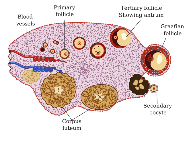
Part (a): The human ovary in section shows primary follicle, secondary oocyte, Graafian follicle and corpus luteum; the gonadotropins FSH and LH drive follicular development and the corpus luteum.
Part (b): An L.S. of the pistil shows the pollen tube entering through the micropyle to the embryo sac (nucellus, antipodals, synergids, micropyle); synergids guide the pollen tube and the micropyle is its entry point.
Part (a)
In a sectional view of the human ovary, the outer cortex (stroma) contains ovarian follicles at successive stages of development:
- (i) Primary follicle — an early follicle with the oocyte surrounded by a layer of granulosa cells.
- (ii) Secondary oocyte — the haploid oocyte within the mature follicle, released at ovulation.
- (iii) Graafian follicle — the fully mature follicle with a fluid-filled antrum, ready to ovulate.
- (iv) Corpus luteum — the yellow endocrine body formed from the ruptured follicle after ovulation.
Hormones influencing follicular development and the corpus luteum: the anterior-pituitary gonadotropins FSH (Follicle-Stimulating Hormone) and LH (Luteinising Hormone). FSH stimulates growth and maturation of the follicles; the mid-cycle LH surge triggers ovulation and the transformation of the ruptured follicle into the corpus luteum, which LH then helps maintain. …
Showing the 12 most recent of 22 on this concept.
- CBSE 2026Set 57/1/11 markMCQQ.Select the option that shows correct statements :(i) Corpus luteum secretes progesterone.(ii) Only FSH attains a peak level in middle of cycle.(iii) LH surge induces rupture of graafian follicle.(iv) Luteal phase of menstrual cycle is also called proliferative phase. Options : (A) Only(ii) is correct. (B)(i) and(iii) are correct. (C) Only(iv) is correct. (D)(ii) and(iv) are correct.
›Reveal solutionSolution
The corpus luteum does secrete progesterone, and the LH surge does trigger ovulation by rupturing the Graafian follicle — making statements (i) and (iii) correct.
The menstrual cycle is a precisely orchestrated hormonal dance, and understanding which hormones do what, and when, is essential to grasping reproductive physiology. Let's examine each statement in turn.
Statement (i): Corpus luteum secretes progesterone.
This is absolutely correct. After ovulation, the ruptured Graafian follicle transforms into the corpus luteum, a temporary endocrine structure in the ovary. The corpus luteum's primary job is to secrete large amounts of progesterone, along with some estrogen. Progesterone prepares the endometrium for implantation by making it thick, vascular, and secretory. If fertilization doesn't occur, the corpus luteum degenerates, progesterone levels drop, and menstruation begins. This is a fundamental fact of the luteal phase.
Statement (ii): Only FSH attains a peak level in middle of cycle.
This is incorrect. While FSH (follicle-stimulating hormone) does rise during the follicular phase to stimulate follicle development, it is not the only hormone that peaks mid-cycle. In fact, the defining hormonal event of mid-cycle is the LH surge — a dramatic spike in luteinizing hormone that occurs around day 14 of a typical 28-day cycle. Both LH and FSH show elevated levels at this time, but the LH surge is far more pronounced and is the direct trigger for ovulation. So the statement that "only FSH" peaks is factually wrong.
Statement (iii): LH surge induces rupture of Graafian follicle.
This is correct. The LH surge is the hormonal signal that causes the mature Graafian follicle to rupture and release the secondary oocyte — the process we call ovulation. Without this surge, ovulation simply does not happen. The LH surge typically occurs about 24–36 hours before ovulation, making it the critical event that marks the transition from the follicular to the luteal phase.
NoteThe LH surge is so reliable that ovulation predictor kits work by detecting elevated LH levels in urine, giving couples a window for conception.
Statement (iv): Luteal phase of menstrual cycle is also called proliferative phase. …
- CBSE 2026Set 57/1/11 markMCQQ.Which connective tissue connects ovary to pelvic wall and uterus ? (A) Tendons (B) Ligaments (C) Cartilage (D) Bone
›Reveal solutionSolution
Ligaments are the connective tissues that anchor the ovary to both the pelvic wall and the uterus, holding the reproductive organs in their proper anatomical positions.
The female reproductive system sits within the pelvic cavity, and like all internal organs, it needs structural support to remain in place during movement, posture changes, and the various physiological processes it undergoes. This is where connective tissues come into play—but not all connective tissues serve the same purpose.
Tendons are specialized connective tissues, but their job is to attach muscle to bone, enabling movement when muscles contract. They have no role in suspending or anchoring organs within body cavities. Cartilage provides cushioning at joints and structural support in places like the nose, ears, and trachea—again, not relevant to holding ovaries in position. Bone forms the rigid framework of the skeleton itself; while the pelvic bones create the cavity that houses the reproductive organs, they don't directly connect to the ovaries.
Ligaments, however, are fibrous connective tissues designed specifically to connect bone to bone or to anchor organs to the body wall. In the female reproductive system, several ligaments perform this anchoring function:
- The ovarian ligament connects each ovary to the lateral wall of the uterus
- The suspensory ligament of the ovary (infundibulopelvic ligament) attaches the ovary to the pelvic wall
- The broad ligament is a fold of peritoneum that helps support the uterus, fallopian tubes, and ovaries …
- CBSE 2026Set A1 markMCQQ.What is the main role of large amount of progesterone secreted by corpus luteum?(a) maintenance of endometrium of uterus(b) maintenance of perimetrium of uterus(c) maintenance of myometrium of uterus(d) None of these
›Reveal solutionSolution
Progesterone from the corpus luteum maintains the uterine endometrium, preparing/keeping it ready for the embryo.
After ovulation, the remaining follicle forms the corpus luteum, which secretes large amounts of progesterone. Progesterone is essential for maintaining the endometrium (the inner lining of the uterus) in a thick, secretory, well-supplied state. This maintained endometrium is necessary for the imp …
- CBSE 2025Set 57/6/11 markMCQQ.Given below are a few statements with respect to spermatogenesis in a human male.(i) Sperms are released from the seminiferous tubules by the process of spermiation.(ii) Spermiogenesis involves the maturation of spermatids into sperms.(iii) Spermatogonia produce spermatids by the process of spermiogenesis.(iv) Meiosis II in secondary spermatocytes results in the formation of four equal haploid spermatids.(v) Primary spermatocyte completes the first meiotic division forming two equal, diploid cells called secondary spermatocytes. Choose the option with all true statements from the given options : (A) (i),(ii) and(iv) (B) (ii),(iii) and(v) (C) (ii),(iv) and(v) (D) (i),(iii) and (iv)
›Reveal solutionSolution
Spermatogenesis involves the transformation of spermatogonia into mature sperms through meiosis and differentiation, culminating in their release from the seminiferous tubules.
Spermatogenesis is the intricate process by which immature male germ cells, called spermatogonia, develop into mature spermatozoa (sperms) within the seminiferous tubules of the testes. This process begins at puberty and continues throughout a male's life, ensuring a continuous supply of gametes for reproduction. It involves a series of mitotic divisions, meiotic divisions, and a final differentiation phase.
Let's break down the key stages and then evaluate the given statements:
- Spermatogonia: These are diploid (2n) undifferentiated germ cells located on the inside wall of the seminiferous tubules. They multiply by mitotic division, increasing their numbers. Some spermatogonia differentiate into primary spermatocytes.
- Primary Spermatocytes: These are also diploid (2n) cells. Each primary spermatocyte then undergoes the first meiotic division (Meiosis I). This is a reductional division, meaning the chromosome number is halved.
- Secondary Spermatocytes: After Meiosis I, each primary spermatocyte forms two equal, haploid (n) cells called secondary spermatocytes. These cells have 23 chromosomes, each with two chromatids.
- Spermatids: Each secondary spermatocyte then rapidly undergoes the second meiotic division (Meiosis II). This is an equational division, similar to mitosis, where sister chromatids separate. As a result, two equal, haploid (n) spermatids are formed from each secondary spermatocyte. Therefore, from one primary spermatocyte, a total of four haploid spermatids are produced.
- Spermiogenesis: This is the process of differentiation where the non-motile, round spermatids are transformed into elongated, motile spermatozoa (sperms). During this phase, the spermatids develop a head, middle piece, and tail.
- Spermiation: After spermiogenesis, the heads of the newly formed sperms become embedded in the Sertoli cells (nurse cells) for nourishment. Once mature, they are finally released from the seminiferous tubules by a process known as spermiation.
Now, let's evaluate each statement:
-
(i) Sperms are released from the seminiferous tubules by the process of spermiation.
- This statement is True. As described above, spermiation is the final step where mature spermatozoa are released from the Sertoli cells into the lumen of the seminiferous tubules.
-
(ii) Spermiogenesis involves the maturation of spermatids into sperms.
- This statement is True. Spermiogenesis is precisely the morphological transformation and differentiation of haploid spermatids into mature, motile spermatozoa.
-
(iii) Spermatogonia produce spermatids by the process of spermiogenesis. …
- CBSE 2025Set A1 markMCQQ.Which part of the ovule is attached to the funicle?(a) Micropyle(b) Hilum(c) Integuments(d) Chalaza
›Reveal solutionSolution
The hilum is the junction between the ovule and its funicle (stalk).
An ovule is attached to the placenta of the ovary by a stalk called the funicle. The point of attachment of the ovule body to the funicle is called the hilum, and it marks the boundary between the funicle and the ovule proper. Other parts named in the options are elsewhere on the ovule: the micropyle is a pore at the opposite end (near the tip) through which the pollen tube usually enters; the i …
- CBSE 2025Set ANNUAL1 markMCQQ.Which hormone is secreted in large amount by the "Corpus luteum"?(a) Testosterone(b) Progesterone(c) Prolactin(d) Thyroxine
›Reveal solutionSolution
After ovulation, the ruptured Graafian follicle becomes the corpus luteum, which mainly secretes progesterone.
Once the ovum is released at ovulation, the remnant of the Graafian follicle transforms into a yellowish endocrine structure called the corpus luteum. Its principal secretion is progesterone, which is essential for maintaining the endometrium of the uterus so that it can support implantation and pregnancy if fertilisation occurs. Testosterone is a …
- CBSE 2025Set ANNUAL1 markQ.Note the relationship between the first two words and fill the missing word.(a) Primary sex organ in male : Testes; Primary sex organ in female : _____.(b) Leydig cells : Androgen; Corpus luteum : _____.
›Reveal solutionSolution
Just as the testis (with its Leydig/interstitial cells secreting androgens) is the male primary sex organ, the ovary is the female primary sex organ, and its corpus luteum secretes the hormone progesterone.
This is a word-relationship (analogy) question testing knowledge of the reproductive organs and their hormone-secreting cells.
- (a) The testis is the primary sex organ (gonad) in the male, responsible for producing sperm (gametogenesis) and male sex hormones. Its exact female counterpart — the primary sex organ (gonad) responsible for producing ova and female sex hormones — is the ovary. …
- CBSE 2025Set ANNUAL1 markQ.Sketch a neat diagram of a typical anatropous ovule.
›Reveal solutionSolution
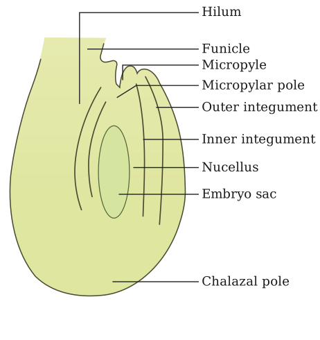
Figure — A hard draw-gate ('Sketch a neat diagram') for a typical anatropous ovule; catalog figure 1-7d is a labelled L An anatropous ovule is completely inverted along its funicle, bringing the micropyle and chalaza close to each other near the base, with the raphe marking the fused stalk.
Diagram (labelled description to draw):
Draw an oval outer body (the ovule, made of the nucellus surrounded by integuments) turned upside-down (180°) relative to its stalk. Label the following parts:
- Funiculus — the stalk attaching the ovule to the placenta.
- Hilum — the point where the funiculus joins the ovule body.
- Raphe — the ridge running along the ovule body, formed because the funiculus is fused lengthwise with the body of the inverted ovule.
- Chalaza — the basal region of the ovule where the integuments and nucellus merge (this ends up close to the hilum in an anatropous ovule).
- Micropyle — the small pore in the integuments through which the pollen tube enters; in an anatropous ovule it lies close to the funiculus, at the same end as the hilum/chalaza. …
- CBSE 2025Set BOTANY1 markMCQQ.Fill in the blank selecting the appropriate one: When pollen tube enters through micropyle into the ovule, it is called ____.(a) porogamy(b) chalazogamy(c) syngamy(d) mesogamy
›Reveal solutionSolution
Pollen-tube entry through the micropyle is porogamy; through the chalaza is chalazogamy; through the integuments/funicle is mesogamy.
After reaching the ovary, the pollen tube grows towards the ovule to deliver the male gametes. Depending on the point of entry into the ovule it is named:
- Porogamy - entry through the micropyle (the normal and most common route).
- Chalazogamy - entry through the chalaza (e.g. Casuarina). …
- CBSE 2024Set E1 markMCQQ.Which of the following secretes large amount of progesterone?(a) Graafian follicle(b) Corpus albicans(c) Corpus luteum(d) Primary follicle
›Reveal solutionSolution
The corpus luteum secretes large amounts of progesterone during the luteal phase.
After ovulation, the remaining parts of the ruptured Graafian follicle transform into a yellowish endocrine structure called the corpus luteum. The corpus luteum secretes large amounts of the hormone progesterone, which is essential for maintenance of the endometrium and for preparing it to receive and support a fertilised ovum (implantation and pregnancy).
…
- CBSE 2024Set ANNUAL1 markMCQQ.Which hormone is secreted by Pituitary gland in human female ?(a) LH(b) FSH(c) Oxytocin(d) All of these
›Reveal solutionSolution
LH, FSH and oxytocin are all pituitary hormones involved in female reproduction.
The anterior pituitary (adenohypophysis) secretes the gonadotropins LH and FSH, which regulate ovarian events (follicular development, ovulation and formation/maintenance of the corpus luteum). The posterior pituitary (neurohypophysis) releases oxytocin, which is important during parturition (uterine contractions) and milk ejection during lactation. Since the pitui …
- CBSE 2024Set ANNUAL1 markMCQQ.Match the following and choose the correct option. Column I Column II
- Ovary A) Androgens
- Corpus luteum B) Human Chorionic gonadotropin
- Leydig cells C) Estrogen + Progesterone
- Placenta D) Progesterone only
(a) 1(A), 2(C), 3(B), 4(D)(b) 1(C), 2(D), 3(A), 4(B)(c) 1(B), 2(A), 3(C), 4(D)(d) 1(A), 2(B), 3(D), 4(C)›Reveal solutionSolution
Ovary → Estrogen + Progesterone, Corpus luteum → Progesterone only, Leydig cells → Androgens, Placenta → hCG, which gives option (b) 1(C), 2(D), 3(A), 4(B).
Matching each source to its principal secretion:
- Ovary — its follicles secrete both estrogen and progesterone, the two chief female sex hormones (option C).
- Corpus luteum — the structure formed from the ruptured Graafian follicle after ovulation; it secretes mainly progesterone to maintain the uterine lining for a possible pregnancy (option D). …
🎓Unlock everything free for 14 days
- ✓Full step-by-step solutions
- ✓Concept-first explanations
- ✓Methods, shortcuts & mistakes
- ✓PYQ mapping + timed mock tests
Full access for 14 days. No credit card required.