Q.Describe the process of megasporogenesis upto fully developed embryo sac formation in an angiosperm.
🔒You're viewing a preview — the full solution, concept, methods & PYQ mapping are locked.
🔒 Start your 14-day free trial to unlock the full solution →Part (a)Concept understanding — Male And Female Gametophyte
Let’s start with something you already know. Think of a plant like a mango tree. It produces flowers, and those flowers eventually turn into mangoes. How does that happen? The flower is the plant’s reproductive organ, and inside it, two tiny but crucial structures are made: the male gametophyte and the female gametophyte. These are not the “male” and “female” parts of the flower themselves (like the stamen and pistil) — they are the microscopic, single-generation cells that actually carry the genetic material to form the next plant.
The everyday intuition
Imagine you are baking a cake. The male gametophyte is like the packet of dry yeast — it’s small, mobile, and needs to be added to the mixture. The female gametophyte is like the bowl of flour, eggs, and sugar — it’s larger, stationary, and provides the environment where the yeast works. Without the yeast reaching the bowl, no cake rises. Without the bowl, the yeast has nothing to act on. In a flower, the male gametophyte (pollen grain) must travel to the female gametophyte (embryo sac) for fertilisation to happen.
The precise meaning
In NCERT biology, the male gametophyte is the pollen grain. It develops inside the anther (the top part of the stamen). A mature pollen grain contains two cells: a tube cell and a generative cell. The tube cell will grow a long tube down the style of the pistil, and the generative cell will divide to form two sperm cells. So the male gametophyte is not the whole stamen — it’s the tiny, single-celled (or two-celled) structure that carries the male genetic material.
The female gametophyte is the embryo sac. It develops inside the ovule, which is located in the ovary of the pistil. The embryo sac is a seven-celled, eight-nucleate structure (though you don’t need to memorise numbers for a prose subject — just know it’s a small, organised sac). It contains the egg cell (the female gamete) and other cells that help in fertilisation and nourishment. The female gametophyte is not the whole pistil — it’s the microscopic sac inside the ovule.
The male gametophyte (pollen grain) is haploid — it has only one set of chromosomes. The female gametophyte (embryo sac) is also haploid. When they fuse during fertilisation, they form a diploid zygote, which grows into the seed. This is why both are called “gametophytes” — they produce gametes (sperm and egg).
Why it matters …
Part (b)Concept understanding — Meiosis And Fertilization
Let’s begin with something you already know: every living thing comes from other living things. A mango tree grows from a seed, and that seed came from a parent mango tree. A puppy is born from its mother. But how does a single cell — a fertilized egg — turn into a whole new person or plant, with the right number of chromosomes and a mix of traits from both parents?
That’s where meiosis and fertilization come in. They are the two halves of the same story: making a new individual while keeping the species stable.
The chromosome problem
Every cell in your body (except sperm and eggs) has 46 chromosomes — 23 from your mother and 23 from your father. That’s the diploid number (2n). If a sperm and an egg each had 46 chromosomes, their fusion would give the baby 92. That would double every generation — impossible. So nature has a neat solution: before sperm and egg meet, each must halve its chromosome count.
That halving is meiosis.
What meiosis does
Meiosis is a special kind of cell division that happens only in the reproductive organs (testes in males, ovaries in females). It takes one diploid cell (2n = 46) and produces four haploid cells (n = 23) — each with one complete set of chromosomes.
Think of it like splitting a deck of cards into two halves, then shuffling each half separately. You end up with four half-decks, each unique.
The process has two rounds of division (meiosis I and meiosis II), but the key point for you is this: meiosis reduces the chromosome number by half and shuffles the genetic material so that each sperm or egg is genetically different from every other.
Why does shuffling matter? Because it creates variation. No two siblings (except identical twins) are exactly alike, and that variety is what allows a species to adapt and survive.
What fertilization does
Fertilization is the opposite of meiosis. It’s the fusion of two haploid cells — a sperm (n = 23) and an egg (n = 23) — to form a single diploid cell called a zygote (2n = 46). That zygote is the first cell of the new individual.
Fertilization restores the full chromosome number. Without meiosis, fertilization would double the count. Without fertilization, meiosis would leave cells with only half the needed chromosomes. The two processes are a matched pair.
Why this matters for you
- Continuity of life: Every human being starts as a single fertilized egg. Meiosis and fertilization are the biological machinery that makes that possible.
- Genetic uniqueness: Because meiosis shuffles chromosomes and fertilization brings together two different sets, every person (except identical twins) has a unique combination of genes. That’s why you look like a blend of both parents but are not identical to either. …
Part (a)
Megasporogenesis is the formation of megaspores from the megaspore mother cell (MMC) inside the nucellus of the ovule.
- A single diploid MMC undergoes meiosis, producing four haploid megaspores in a linear tetrad.
- In most angiosperms three megaspores degenerate; only the functional megaspore (usually the one at the chalazal end) survives.
- The functional megaspore now forms the female gametophyte (embryo sac) by megagametogenesis: its nucleus undergoes three free-nuclear mitotic divisions giving 8 nuclei (4 at each pole). …
Part (a): the MMC undergoes meiosis → functional megaspore → three mitoses → 7-celled, 8-nucleate embryo sac.
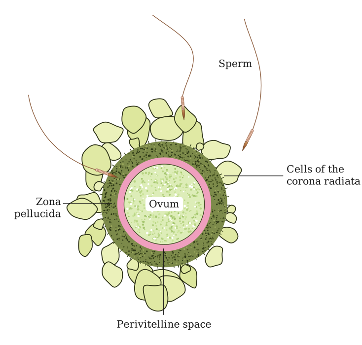
Part (b): the sperm nearest the ovum reaches first; the acrosome's enzymes allow entry, which triggers the cortical reaction and completion of meiosis-II, all in the ampulla of the fallopian tube.
Part (a)
The female gametophyte of an angiosperm develops inside the ovule, which lies within the ovary.
Megasporogenesis. A single diploid cell in the nucellus, the megaspore mother cell (MMC), enlarges and undergoes meiosis (a reduction division). This produces four haploid megaspores, usually arranged in a linear tetrad. In the great majority of flowering plants three of these megaspores degenerate and only one, the functional megaspore (typically the one farthest from the micropyle, at the chalazal end), survives. This step — MMC to functional megaspore — is megasporogenesis.
Megagametogenesis (embryo sac formation). The functional megaspore enlarges and its nucleus divides by three successive free-nuclear mitotic divisions:
- first mitosis → 2 nuclei, which move to opposite poles;
- second mitosis → 4 nuclei (2 at each pole);
- third mitosis → 8 nuclei (4 at each pole).
Cell walls now lay down around these nuclei to give the characteristic 7-celled, 8-nucleate structure (the Polygonum type, the commonest in angiosperms):
- Micropylar end — an egg apparatus of one egg cell flanked by two synergids (the synergids bear the filiform apparatus that guides the pollen tube).
- Chalazal end — three antipodal cells.
- Centre — a large central cell whose two polar nuclei later fuse to give the diploid secondary nucleus. …
Showing the 12 most recent of 86 on this concept.
- CBSE 2026Set 57/2/11 markMCQQ.Given below are stages of embryo sac formation from megaspore mother cell. Identify the correct option for the cell division marked a, b, c and d. (A) a – Meiosis-I, b – No division, c – Mitosis, d – Meiosis-II (B) a – Meiosis-I, b – Meiosis-II, c – No division, d - Mitosis (C) a – Mitosis, b – Meiosis-I, c – Meiosis-II, d – No division (D) a – No division, b – Mitosis, c – Meiosis-I, d – Meiosis-II
›Reveal solutionSolution
The megaspore mother cell undergoes meiosis (two divisions) to produce four haploid megaspores, of which only one survives; that functional megaspore then undergoes three mitotic divisions to form the eight-nucleate embryo sac — so the correct sequence is Meiosis-I, Meiosis-II, No division, Mitosis.
To understand this question, you need to picture the sequence of events inside the ovule of a flowering plant. The story begins with a single, large cell called the megaspore mother cell (MMC) . This cell is diploid (2n) and is the starting point for forming the female gametophyte — the embryo sac.
The MMC does not simply grow or divide by mitosis. Instead, it enters meiosis, a special type of cell division that reduces the chromosome number by half. Meiosis happens in two consecutive stages: Meiosis-I and Meiosis-II. So the very first division (marked 'a' in your diagram) is Meiosis-I. The second division (marked 'b') is Meiosis-II. After these two divisions, the original single MMC has produced four haploid (n) cells — these are called megaspores.
Now here is a critical twist. In most flowering plants, three of these four megaspores degenerate and die. Only one of them — usually the one farthest from the micropyle (the opening of the ovule) — remains functional. So after the four megaspores are formed, there is a stage where no division occurs (marked 'c' in your diagram). Instead, three cells simply break down, leaving just one functional megaspore.
That single functional megaspore is the starting point for the actual embryo sac. It is haploid and now needs to grow into a structure that contains the egg cell and other supporting cells. How does it do that? It undergoes three rounds of mitotic divisions (marked 'd'). Mitosis ensures that all the resulting nuclei remain haploid and genetically identical. The first mitosis produces two nuclei, the second produces four, and the third produces eight nuclei. These eight nuclei then arrange themselves into the typical seven-celled, eight-nucleate embryo sac (with the egg apparatus, central cell, and antipodal cells).
So, mapping this to the labels in your question:
- a = Meiosis-I (first reduction division)
- b = Meiosis-II (second division, producing four megaspores)
- c = No division (three megaspores degenerate) …
- CBSE 2026Set A1 markMCQQ.Which of the following is one of the most resistant organic materials?(a) Pectin(b) Cellulose(c) Sporopollenin(d) Chitin
›Reveal solutionSolution
Sporopollenin forms the pollen exine and is among the most resistant organic materials known.
The hard outer wall (exine) of a pollen grain is made of sporopollenin. It can withstand high temperatures and strong acids and alkalis, and no enzyme is known that can degrade it. This extreme resistance is wh …
- CBSE 2026Set A1 markMCQQ.Which of the following cells is present in pollen grain of angiospermic plants?(a) Vegetative cell(b) Generative cell(c) Antipodal cell(d) Both (A) and (B)
›Reveal solutionSolution
An angiosperm pollen grain (male gametophyte) contains a vegetative cell and a generative cell.
When a pollen grain is shed, it is usually at the 2-celled stage. It contains a large vegetative (tube) cell with abundant food reserves, and a small generative cell floating within its cytoplasm. The generative cell later divides to form two male ga …
- CBSE 2026Set A1 markMCQQ.Which of the following plays an important role in guiding the pollen tube into the synergids?(a) Antipodals(b) Filiform apparatus(c) Central cell(d) Micropyle
›Reveal solutionSolution
The filiform apparatus of the synergids guides the entering pollen tube into a synergid.
At the micropylar end of the embryo sac, the synergids bear special cellular thickenings called the filiform apparatus. This structure guides the pollen tube by directing its entry into one of the synergids. Once inside, the pollen tube bursts and releases the …
- CBSE 2026Set ANNUAL1 markQ.What does 'A' and 'B' represent in the above mentioned diagram of the mature embryosac? Write name for each.
›Reveal solutionSolution
In the mature 7-celled, 8-nucleate embryo sac, A marks the two polar nuclei of the central cell and B marks the three-celled egg apparatus (2 synergids + 1 egg cell) at the micropylar end.
A typical angiosperm embryo sac at maturity has 7 cells and 8 nuclei, organised as: 3 antipodal cells at the chalazal end; a large central cell containing 2 polar nuclei (which fuse with a male gamete during triple fusion to form the triploid endosperm); and, at the micropylar end, the egg apparatus consisting of 1 egg cell flanked by 2 synergid cells. The synergids bear a special thickened structure at their micropylar tip called the filiform apparatus, which guides entry of the pollen tube into the embryo sac. In the given figure, going from the chalazal end (top, unlabelled antipodal cells) toward the micropylar end (bottom, where the filiform a …
- CBSE 2026Set ANNUAL1 markMCQQ.Male gametes in angiosperms are formed by the division of(a) Vegetative cell(b) Generative cell(c) Microspore mother cell(d) Megaspore mother cell
›Reveal solutionSolution
The pollen grain's generative cell divides mitotically to form two non-motile male gametes that are delivered through the pollen tube.
A mature pollen grain in angiosperms is typically 2-celled at shedding: a larger vegetative (tube) cell and a smaller generative cell, floating within the vegetative cell's cytoplasm.
- The vegetative cell has abundant food reserves and, upon germination on the stigma, forms the pollen tube that grows down through the style. …
- CBSE 2026Set ANNUAL1 markQ.Where is filiform apparatus located? What is the role of filiform apparatus?
›Reveal solutionSolution
The filiform apparatus is a set of finger-like cell-wall thickenings at the micropylar end of each synergid, and it functions to guide the entry of the pollen tube into the embryo sac.
Location: Within the mature embryo sac of an angiosperm ovule, two synergid cells flank the egg cell at the micropylar end (together forming the egg apparatus). Each synergid has specialised, finger-like thickenings of its cell wall at its micropylar tip called the filiform apparatus.
…
- CBSE 2026Set ANNUAL1 markQ.What is the number of chromosomes in spermatogonial cells and spermatids of a human?
›Reveal solutionSolution
Spermatogonia are diploid stem cells (46 chromosomes) inside the seminiferous tubules; after meiosis they give rise to haploid spermatids (23 chromosomes), which then differentiate into sperm.
In the human testis, spermatogonial cells are diploid (2n = 46 chromosomes) stem cells located along the inner wall of the seminiferous tubules. Some spermatogonia divide mitotically to maintain their own numbers, while others (type B spermatogonia) enter meiosis as primary spermatocytes.
…
- CBSE 2026Set ANNUAL1 markMCQQ.Pollen grains retain viability for months in plant belonging to different families given below :(i) Solanaceae(ii) Leguminosae(iii) Gramineae(iv) Rosaceae(v) Liliaceae Correct option is :(a) (i),(ii) &(v)(b) (i),(ii) &(iv)(c) (ii), (iv),(v)(d) (i), (iii), (v)
›Reveal solutionSolution
Pollen viability varies enormously by species — from 30 minutes in some cereals to months in members of Solanaceae, Leguminosae and Rosaceae.
Pollen grain lifespan depends on temperature and humidity. In many members of the family Poaceae (Gramineae) pollen viability is lost within 30 minutes of release, while in some members of Solanaceae, Leguminosae, and Rosaceae, pollen grains maintain viability for …
- CBSE 2026Set ANNUAL1 markMCQQ.The structure of bilobed anther consist of :(a) 2 thecae, 2 sporangia(b) 4 thecae, 4 sporangia(c) 4 thecae, 2 sporangia(d) 2 thecae, 4 sporangia
›Reveal solutionSolution
A typical anther is bilobed and dithecous: two lobes (thecae), each theca bearing two microsporangia (pollen sacs) — four microsporangia in total.
A mature anther consists of two lobes joined by a sterile connective. Each lobe is called a theca, so a bilobed anther has 2 thecae, making it 'dithecous'. Within each theca lie two elongated sac-like structures called microsporangia (pollen sacs), running almost the full length of the anther. So each theca contributes 2 microsporangia, giving …
- CBSE 2026Set ANNUAL1 markMCQQ.Egg apparatus is made up of :(a) 2 synergid cells + 2 egg cells(b) 1 synergid cell + 2 egg cells(c) 2 synergid cells + 1 egg cell(d) 1 synergid cell + 1 egg cell
›Reveal solutionSolution
The egg apparatus, sitting at the micropylar end of the embryo sac, is made up of 2 synergid cells + 1 egg cell.
The mature female gametophyte (embryo sac) of a flowering plant is typically 7-celled and 8-nucleate. At its micropylar end lies a group of three cells called the egg apparatus, consisting of:
- 1 egg cell (oosphere) — the female gamete, located in the centre, flanked by the two synergids. …
- CBSE 2026Set ANNUAL1 markMCQQ.During fertilization, which cell fuses with male gamete to form zygote?(a) Egg Cell(b) Polar Cell(c) Synergid(d) Antipodal Cell
›Reveal solutionSolution
Syngamy (fusion of the egg cell with one male gamete) forms the diploid zygote; the other male gamete fuses with the polar nuclei (triple fusion) to form the endosperm — together this is double fertilization.
In the mature embryo sac, the egg apparatus consists of one egg cell flanked by two synergids, with three antipodal cells at the chalazal end and two polar nuclei in the central cell. When the pollen tube discharges its two male gametes into a synergid:
- One male gamete fuses with the egg cell → forms the diploid zygote (syngamy). …
🎓Unlock everything free for 14 days
- ✓Full step-by-step solutions
- ✓Concept-first explanations
- ✓Methods, shortcuts & mistakes
- ✓PYQ mapping + timed mock tests
Full access for 14 days. No credit card required.