Biology · Ch 9 — Control and Co-ordination
Spinal Cord
Spinal Cord
B. Spinal Cord : The spinal cord is the lower continuation of the medulla oblongata, running inside the vertebral column's neural canal and, like the brain, protected by bony covering and the same three meninges (dura, arachnoid and pia mater), with CSF secreted by the pia mater forming a fluid cushion around it and inside its central canal. Externally it is a long cylindrical rod, 42-45 cm long and 2.0-2.5 cm broad, broadest at its anterior end and tapering posteriorly first into the conus medullaris (around vertebral level L1-L2) and then into a thread-like filum terminale; it also shows two swellings, cervical and lumbar, where large numbers of spinal nerves emerge. Thirty-one pairs of spinal nerves arise from its lateral sides, concentrated near the cervical and lumbar swellings and around the conus medullaris; the bundle of nerves plus the filum terminale trailing from the cord's lower end resembles a horse's tail and is called the cauda equina.
T. S. of spinal cord : In cross-section, the spinal cord is dorsoventrally flattened by a deep, narrow posterior median fissure and a shallow, broad anterior median fissure, which incompletely divide it into right and left halves around a central canal. Butterfly (H)-shaped grey matter occupies the centre, divided by the fissures into six horns (dorsal, lateral and ventral, on each side), while white matter occupies the outside, divided into six columns/funiculi (again dorsal, lateral and ventral). The dorsal and ventral horns extend out of the cord as the dorsal root and ventral root respectively; the dorsal root connects to the dorsal root ganglion (just outside the cord), which holds the cell bodies of unipolar sensory neurons. Inside the grey matter, association (inter-)neurons receive sensory input, integrate it, and direct the response to motor neurons in the ventral horn, while neurons of the autonomic nervous system sit in the lateral horn and leave via the ventral root. The white matter's myelinated fibre bundles form ascending tracts (carrying sensory impulses from cord to brain, in the dorsal funiculi) and descending tracts (carrying motor impulses from brain to the lateral and ventral funiculi).
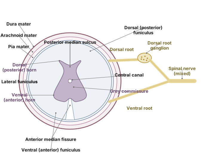
Drawn by us to help you understand the concept clearly, and verified to make sure it's accurate. For exams, practice from your textbook's own diagram.
What this figure shows. A cross-section of the spinal cord showing the deep, narrow posterior median fissure and the shallow, broad anterior median fissure incompletely dividing it into two sides; centrally, H-shaped (butterfly-shaped) grey matter with dorsal, lateral and ventral horns surrounding the central canal (lined by ependyma); and, external to the grey matter, white matter from which a dorsal (sensory) root and a ventral (motor) root emerge on each side.
9.11: T. S. of spinal cord. …
…