Biology · Ch 9 — Control and Co-ordination
Spinal Nerves
Spinal Nerves
The peripheral nervous system connects the CNS to receptors and effectors throughout the body, and its nerves are classically split by connection point into cranial nerves (linked to the brain, covered above) and spinal nerves (linked to the spinal cord); more recent classification instead splits the PNS functionally into the somatic nervous system (relaying CNS impulses to skeletal/voluntary muscle) and the autonomic nervous system (covered separately below).
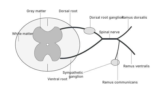
Spinal Nerves : Thirty-one pairs of spinal nerves, all of the mixed type, arise from the spinal cord in five named groups by vertebral region -- 8 cervical (C1-C8, from the neck), 12 thoracic (T1-T12, from the thorax), 5 lumbar (L1-L5, from the abdomen), 5 sacral (S1-S5, from the pelvis) and 1 coccygeal (Co1, from the coccyx) -- and provide two-way communication between the spinal cord and the neck, trunk and limbs. Each spinal nerve forms inside the vertebral canal from two roots: the posterior/dorsal root, which is sensory and receives fibres from the dorsal root ganglion (holding the sensory-neuron cell bodies), and the anterior/ventral root, which gives out motor fibres; together these dorsal-sensory and ventral-motor components join into one mixed spinal nerve, which then emerges through the intervertebral foramen and immediately splits into three branches -- the ramus dorsalis (to skin and muscles of the dorsal side), the ramus ventralis (the largest of the three, supplying organs and muscles on the lateral and anterior side), and the ramus communicans (the smallest, present from the first thoracic to the third lumbar spinal nerve, which joins the sympathetic ganglia of the au …
Cervical: 8 pairs (C1-C8), origin -- neck.
Thoracic: 12 pairs (T1-T12), origin -- thorax.
Lumbar: 5 pairs (L1-L5), origin -- abdomen.
Sacral: 5 pairs (S1-S5), origin -- pelvis. …
Drawn by us to help you understand the concept clearly, and verified to make sure it's accurate. For exams, practice from your textbook's own diagram.
What this figure shows. A diagram showing how a typical spinal nerve is built: a dorsal root carrying sensory fibres (bearing a dorsal root ganglion of sensory-neuron cell bodies) and a ventral root carrying motor fibres emerge from the grey and white matter of the spinal cord and join just outside the vertebral canal to form one mixed spinal nerve, which then splits into a ramus dorsalis, a larger ramus ventralis, and a small ramus communicans that connects to …