Biology · Ch 4 — Molecular Basis of Inheritance
Packaging in Eukaryotes
Packaging in Eukaryotes
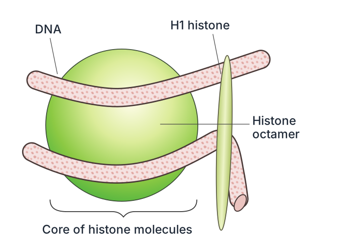
Packaging in Eukaryotes: Eukaryotic cells have a well-organised nucleus with a nuclear membrane, a nucleolus, and thread-like chromosomes in which DNA is associated with histone and non-histone proteins (as reported by R. Kornberg in 1974). Histones are rich in the basic amino acids lysine and arginine, which carry a positive charge; this lets them bind tightly to the negatively charged DNA backbone. Eight histone molecules -- two each of H2A, H2B, H3 and H4 -- assemble into a histone octamer, and about 200 base pairs of DNA wrap around each octamer (roughly 146 bp in about 1(3/4) turns), with the rest forming linker DNA between one nucleosome and the next; a further histone, H1, binds where the DNA enters and leaves the nucleosome (Fig. 4.5).
Drawn by us to help you understand the concept clearly, and verified to make sure it's accurate. For exams, practice from your textbook's own diagram.
What this figure shows. A magnified view of a single nucleosome: a core histone octamer (two molecules each of H2A, H2B, H3 and H4) with roughly 146 base pairs of the negatively charged DNA double helix wound around it in about 1(3/4) turns, and an H1 histone molecule seated where the DNA strand enters and leaves the core, clamping the wrapped DNA in place; a stretch of link …
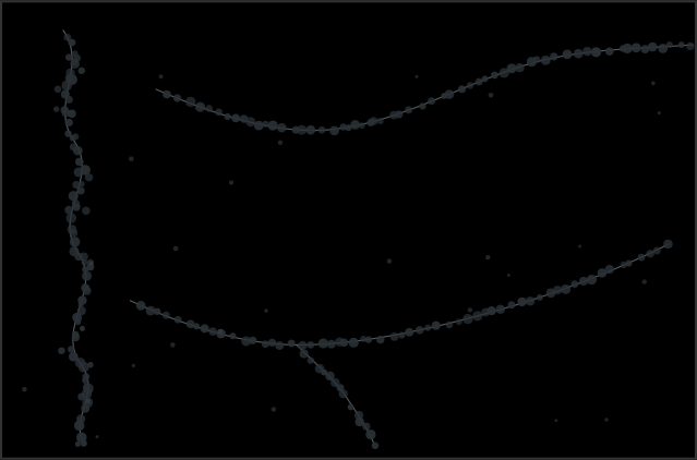
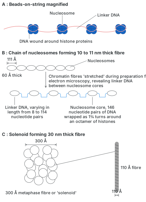
Under an electron microscope, this chain of nucleosomes on linker DNA looks like 'beads-on-string' (Fig. 4.6).
Drawn by us to help you understand the concept clearly, and verified to make sure it's accurate. For exams, practice from your textbook's own diagram.
What this figure shows. A three-part electron-microscope-style illustration of chromatin at increasing magnification: panel A is an enlarged 'beads-on-a-string' view of individual nucleosome beads joined by linker DNA of variable length (about 8-114 nucleotide pairs); panel B shows a chain of such nucleosomes forming a roughly 10-11 nm thick fibre; and panel C shows that fibre further coiling into a thicker roughly 30 nm 'solenoid' fibre, illustrating the first levels of chro …
…
Drawn by us to help you understand the concept clearly, and verified to make sure it's accurate. For exams, practice from your textbook's own diagram.
What this figure shows. A schematic showing nucleosomes (DNA wound around histone protein cores, connected by linker DNA) coiling together, about six nucleosomes per turn, into a helical 30 nm solenoid structure resembling a coiled telephone cord -- illustrating the packaging level above the simple beads-on-a-string chromatin fibre, on the way to the fully …