Biology · Ch 4 — Molecular Basis of Inheritance
Translation
Translation
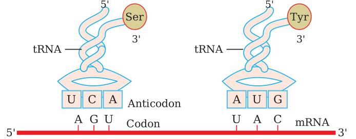
t-RNA - the adapter molecule: Scientists reasoned that there must be a mechanism by which tRNA reads the codon and at the same time binds the amino acid, since an amino acid has no special capacity to read a codon; tRNA is therefore considered an adapter molecule, a role understood much later. Folded by internal base pairing into a two-dimensional cloverleaf shape (and an inverted-L shape in three dimensions), each tRNA has an anticodon loop whose three bases pair with a specific mRNA codon, and a single-stranded 3' end bearing an unpaired CCA sequence, to which one specific amino acid is attached. A separate tRNA exists for each amino acid (with a special initiator tRNA specific to methionine), and, since there is no amino acid corresponding to a stop signal, no tRNA recognises the stop codons.
Drawn by us to help you understand the concept clearly, and verified to make sure it's accurate. For exams, practice from your textbook's own diagram.
What this figure shows. A cloverleaf (two-dimensional) diagram of a transfer RNA molecule folded by intramolecular base pairing into stem-and-loop arms, highlighting the anticodon loop at the bottom (shown here pairing with a codon) and the single-stranded 3' amino-acid-acceptor end bearing the unpaired CCA sequence, to which a specific amino acid (glutamic acid, in this example) is attached -- illustrating why tRNA is described as an adapter that links a …
Can you tell?
Why is tRNA called an adapter molecule?
Can you recall?
- Name the different types of RNAs.
- Name the site of protein synthesis.
- Which molecule carries the information for protein synthesis from the gene?
- Which molecule carries the amino acid from the cytoplasm to the ribosome?
B. Translation - protein synthesis : Translation is the mechanism in which the codons of mRNA are translated and specific amino acids, in sequence, form a polypeptide on the ribosomes. All types of proteins are synthesised by the cell within itself (intracellularly). Translation requires amino acids, mRNA, tRNA, ribosomes, ATP, Mg++ ions, enzymes, and elongation, translocation and release factors:
i. Amino acids are the raw material of protein synthesis; about 20 different amino acids form proteins, and they are available in the cytoplasm.
ii. DNA controls the synthesis of proteins with amino acids in a specific sequence; this control works through the transcription of mRNA, and the genetic code is specific for each amino acid.
iii. RNAs serve as the intermediate molecules between DNA and protein.
iv. Ribosomes are the site of protein synthesis. Each ribosome consists of a large and a small subunit, which lie separately in the cytoplasm and associate only during protein synthesis, with the help of Mg++ ions.
A ribosome has one binding site for mRNA and three for tRNA: the P site (peptidyl-tRNA site), the A site (aminoacyl-tRNA site) and the E site (exit site). Only the first tRNA-amino acid complex enters the P site directly. In eukaryotes a groove between the two subunits protects the polypeptide chain from cellular enzymes and the mRNA from nucleases.
Mechanism of translation (synthesis of the polypeptide chain) : It involves three steps: i. initiation, ii. elongation, iii. termination.
1. Initiation of Polypeptide chain :
a. Activation of amino acids, which needs ATP, is essential before translation begins. The small subunit of the ribosome binds to the mRNA at its 5' end. The initiator codon AUG on the mRNA starts translation; the initiator tRNA binds to it by its anticodon (UAC) through hydrogen bonds and carries the activated amino acid methionine (in eukaryotes) or formyl-methionine (in prokaryotes).
b. The large subunit of the ribosome now joins the small subunit, which requires Mg++ ions.
c. The initiator charged tRNA (with methionine) occupies the P site of the ribosome, and the A site is vacant.
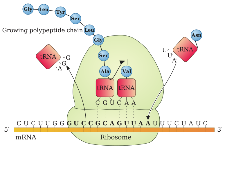
2. Elongation of polypeptide chain : Activated amino acids are added one by one to the first amino acid (methionine). An amino acid is activated with energy from ATP and binds to the amino-acid binding site of its tRNA, forming a tRNA-amino acid complex. Each addition is a three-step cycle:
a. Codon recognition - the aminoacyl-tRNA enters the ribosome at the A site and its anticodon binds the codon by hydrogen bonds.
b. The amino acid on the initiator tRNA at the P site and the amino acid on the tRNA at the A site join by a peptide bond, with the enzyme ribozyme as catalyst; the first tRNA at the P site is then kicked off.
c. Translocation - the tRNA at the A site, now carrying a dipeptide, moves to the P site; both ribosomal subunits move along relative to the tRNA and mRNA. The tRNA with the dipeptide sits at the P site, leaving the A site vacant for the next charged tRNA, while the first uncharged tRNA is discharged from the E site. This repeats as amino acids are added to the polypeptide; a peptide bond forms in less than 0.1 second. The third charged tRNA arrives at the A site, anticodon and codon bind, the peptide bond forms and the second tRNA is discharged from the P site to the E site and leaves the ribosome. The events - arrival of the tRNA-amino acid complex, peptide-bond formation, ribosomal translocation and removal of the previous tRNA - are repeated, and as the ribosome moves over the mRNA all its codons are exposed one by one for translation. …
Drawn by us to help you understand the concept clearly, and verified to make sure it's accurate. For exams, practice from your textbook's own diagram.
What this figure shows. A three-panel diagram of polypeptide synthesis on a ribosome: panel (a) initiation, showing the small ribosomal subunit bound to mRNA with the initiator tRNA in the P site and the large subunit about to join; panel (b) elongation, showing a charged tRNA entering the vacant A site, peptide-bond formation with the growing chain, and translocation of the ribosome that shifts tRNAs from the A to the P to the E site as the discharged tRNA leaves; and panel (c) termination, showing the ribosome positioned over a stop codon, the completed polypeptide chain and the last tRNA bein …