Biology · Ch 2 — Reproduction in Lower and Higher Animals
Cleavage
Cleavage
Cleavage : It is the process of early, rapid mitotic division of the zygote, converting it into a hollow, multicellular blastula; unlike ordinary cell division, cleavage does not involve growth of the daughter cells. The cells produced by cleavage are called blastomeres. Because there is no growth phase between successive cleavages, the size of the blastomeres becomes progressively smaller with each division; as their size decreases, their metabolic rate rises, so successive cleavages actually occur faster than the ones before — a pace that demands rapid DNA replication and high oxygen consumption.
Process of cleavage : In humans, cleavage is holoblastic, meaning the whole zygote is divided at each cleavage. The cleavage planes may run longitudinally (meridional) or transversely (equatorial/horizontal), and cleavage is radial and indeterminate — the eventual fate of each blastomere is not fixed in advance. The first cleavage in the zygote is meridional, occurring about 30 hours after fertilization, and divides the zygote longitudinally into two blastomeres, one slightly larger than the other. The second cleavage is also longitudinal, but at a right angle to the first, and occurs roughly 30 hours after the first cleavage. The third cleavage is horizontal, producing, by this point, an 8-cell embryo. All the while, the successive cleavages push the developing embryo steadily towards the uterus. By the end of the fourth day after fertilization, the embryo is a solid ball of 16–32 cells, externally resembling a mulberry — this stage is therefore called the morula. The morula shows two types of cells: smaller, clearer cells on the outside, and an inner cell mass of larger cells; the cells remain tightly compact, and until the morula stage the zo …
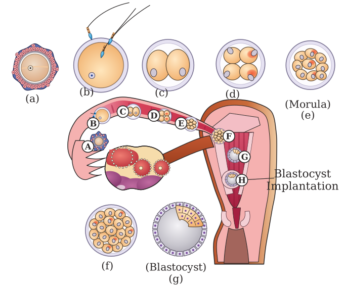
Drawn by us to help you understand the concept clearly, and verified to make sure it's accurate. For exams, practice from your textbook's own diagram.
What this figure shows. The stages the textbook's Fig. 2.14 lettered A to F, here laid along the fallopian tube: the fertilized ovum, the 2-cell stage (about 30 hours), the 4-cell and 8-cell stages, the solid morula, and the blastocyst with its trophoblast, blastocyst cavity and inner cell mass …