Biology · Ch 1 — Reproduction in Lower and Higher Plants
Development of Embryo
Development of Embryo
Embryogenesis, the development of the zygote into a fully formed embryo, begins only after a certain amount of endosperm has already formed around it, ensuring the developing embryo has a nutritive tissue to draw upon from the outset. The embryo itself develops at the micropylar end of the embryo sac.
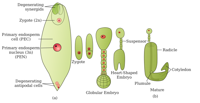
The zygote's first division produces a two-celled proembryo made of a larger cell nearer the micropyle, the basal or suspensor initial cell, and a smaller cell nearer the chalaza, the terminal or embryonal initial cell. The suspensor initial then divides transversely, in a single plane, to build up a filamentous suspensor of about six to ten cells, whose job is to physically push the developing embryo deeper into the nutritive endosperm. The uppermost suspensor cell (nearest the micropyle) swells and functions as an absorptive haustorium, while the lowermost suspensor cell, immediately adjacent to the true embryo, is called the hypophysis and later contributes to the root.
Meanwhile, the embryonal initial cell undergoes three successive mitotic divisions, in mutually perpendicular planes, to form an eight-celled stage called the octant. The lower tier of four octant cells goes on to form the hypocotyl and radicle (embryonic root axis), while the upper tier of four cells forms the plumule (embryonic shoot) and one or two cotyledons (seed leaves); the hypophysis, by further division, contributes the remainder of the radicle and the protective root cap. As development proceeds, the upper-tier cells divide in multiple planes to become heart-shaped, differentiating into two lateral cotyledons flanking a terminal plumule; continued enlargement of the hypocotyl and cotyledons then curves the whole embryo, giving the mature dicot embryo its characteristic horseshoe shape (illustrated in Capsella, the classic dicot embryogenesis model).
Drawn by us to help you understand the concept clearly, and verified to make sure it's accurate. For exams, practice from your textbook's own diagram.
What this figure shows. Panel (a) is the embryo sac just after double fertilization: the diploid zygote (2n) sits at the micropylar end beside the degenerating synergids, the large primary endosperm cell holds the triploid primary endosperm nucleus (3n), and the antipodal cells at the chalazal end are degenerating. Panel (b) follows the zygote through embryogeny, the sequence the textbook's Fig. 1.15 draws for Capsella (stages A to H): the zygote (oospore) divides into a two-celled proembryo; the basal cell builds the filamentous suspensor while the terminal cell forms the globular embryo (the octant stage and its derivatives); the embryo becomes heart-shaped as the two cotyledons start to bulge; and the mature embryo curves round with its radicle, plumule and two cotyledons, by which time the endosperm has been …
…
Drawn by us to help you understand the concept clearly, and verified to make sure it's accurate. For exams, practice from your textbook's own diagram.
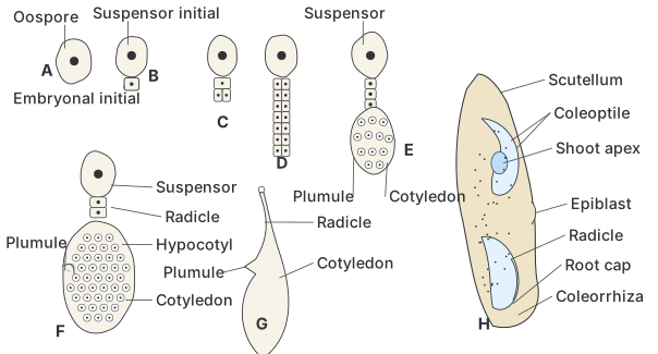
What this figure shows. This diagram traces the development of a monocot (grass-type) embryo, showing how, unlike the dicot pattern, a single shield-shaped cotyledon called the scutellum occupies the terminal position while the plumule develops laterally; the diagram labels the protective coleoptile sheath around the plumule and the coleorhiza sheath around the radicle, along with the shoot apex …