Biology · Ch 1 — Reproduction in Lower and Higher Plants
Megasporogenesis and Development of Female Gametophyte
Megasporogenesis and Development of Female Gametophyte
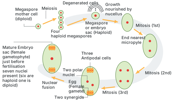
Megasporogenesis is the process by which a diploid megaspore mother cell (MMC), which differentiates within the nucellus roughly at its centre but displaced toward the micropylar end of the ovule, undergoes meiosis to produce a linear tetrad of four haploid megaspores. Of these four, the three megaspores nearer the micropylar end degenerate and abort, while only the single megaspore lying towards the centre of the nucellus (the chalazal-most of the tetrad) survives and becomes functional. This surviving cell becomes the first cell of the female gametophyte (embryo sac).
Development of female gametophyte. The functional megaspore then develops into the mature embryo sac through three successive rounds of free-nuclear mitotic division (i.e., nuclear division without immediate wall formation), producing a total of eight nuclei, four located at each pole of the developing sac. Because this whole embryo sac traces back to a single functional megaspore, this pattern of development is termed monosporic development, and because it takes place entirely within the confines of the megaspore/nucellus, angiosperm female gametophyte development is described as endosporous.
Once the eight nuclei are in place, cell walls partition most of them into a seven-celled structure (the eighth nucleus arising from fusion, described below), organised as follows:
- Three nuclei at the micropylar end form the egg apparatus: one large, central haploid egg cell (the female gamete) flanked by two smaller haploid synergid cells. Each synergid bears finger-like cytoplasmic projections called the filiform apparatus, which chemically guide the growing pollen tube toward the egg.
- Three nuclei at the chalazal end remain as separate cells called antipodal cells, which play a nutritive/supportive role and eventually degenerate.
- One nucleus migrating in from each pole, the two polar nuclei, come to lie together in the large central cell and fuse (just before fertilization) to form a diploid secondary nucleus (also called the definitive nucleus). …
Drawn by us to help you understand the concept clearly, and verified to make sure it's accurate. For exams, practice from your textbook's own diagram.
What this figure shows. This is a step-by-step diagram of female gametophyte (embryo sac) development from the diploid megaspore mother cell. It traces meiosis producing four haploid megaspores, of which the three nearer the micropyle degenerate while the functional one nearest the centre survives, followed by three successive free-nuclear mitotic divisions of the surviving megaspore that generate eight nuclei, and finally wall formation that organises these into the mature seven-celled embryo sac, showing the egg cell with its two synergids at the micropylar end, the three antip …