Biology · Ch 1 — Reproduction in Lower and Higher Plants
Structure of Anatropous Ovule
Structure of Anatropous Ovule
The gynoecium (pistil) is the female reproductive whorl of the flower, and each individual unit of the gynoecium is called a carpel, or megasporophyll. A flower whose gynoecium consists of several free, unfused carpels is described as apocarpous (as in Michelia), whereas one whose carpels are fused together into a single compound structure is syncarpous (as in brinjal). A typical carpel is differentiated into three regions: the swollen basal ovary, which houses the ovule(s); the slender style connecting the ovary to the stigma; and the stigma at the tip, which receives pollen. The number of ovules housed within the ovary varies widely between species -- paddy, wheat, and mango carry only a single ovule (uniovulate), while tomato and lady's finger carry many (multiovulate).
Each ovule develops inside the ovary, attached to the placental tissue by a short stalk called the funiculus (or funicle); the point where this stalk meets the main body of the ovule is called the hilum. The commonest ovule type in angiosperms is the anatropous ovule, in which the ovule has become inverted during development so that its micropyle (the narrow opening at its apex, and the eventual entry point for the pollen tube) points downward, lying close beside the funiculus. Internally, the ovule is built around a central mass of parenchymatous tissue, the nucellus, which is itself surrounded by two protective layers, the outer integument and the inner integument. The base of the ovule, directly opposite the micropyle, is called the chalaza. Embedded within the nucellus lies the embryo sac, the oval, multicellular female gametophyte, whose own internal structure and development are described under megasporogenesis.
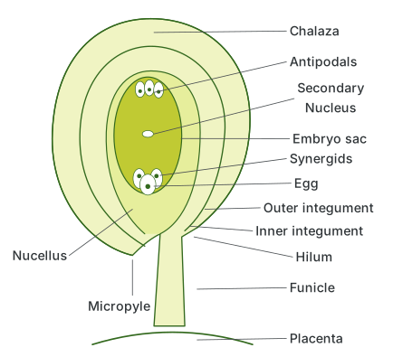
Drawn by us to help you understand the concept clearly, and verified to make sure it's accurate. For exams, practice from your textbook's own diagram.
What this figure shows. This is a labelled longitudinal-section diagram of a typical anatropous ovule. It shows the ovule attached by its funicle to the placenta, with the point of attachment called the hilum; the outer and inner integuments surrounding the central nucellus; the micropyle positioned close to the funicle because the ovule is inverted; the chalaza at the base opposite the micropyle; and, embedded within the nucellus, the embryo sac containing the egg cell, two flanking syner …
…