Biology · Ch 1 — Reproduction in Lower and Higher Plants
Development of Endosperm
Development of Endosperm
Following triple fusion, the resulting triploid primary endosperm nucleus divides repeatedly by mitosis to build up a nutritive tissue called the endosperm, which develops within the ovule alongside, and typically slightly ahead of, the developing embryo, providing it with a ready food supply. As endosperm formation proceeds, the other residual cells of the original embryo sac (the synergids and antipodals) gradually disorganise and are absorbed.
There are three types of endosperm on the basis of the mode of development: i. Nuclear type, ii. Cellular type, iii. Helobial type.
a. Nuclear Type : The most widespread pattern, found in roughly 161 angiosperm families. The primary endosperm nucleus divides mitotically again and again without any accompanying wall formation, producing a large number of free nuclei suspended in a common cytoplasm. A large central vacuole then forms, pushing all these free nuclei out toward the periphery of the cell; only later do walls begin to form between the nuclei, converting the tissue into a proper multicellular endosperm. In several plants, wheat, sunflower, and coconut among them, this wall formation never becomes fully complete; the coconut, distinctively, ends up with a solid multicellular endosperm in its outer region while the central region stays free-nuclear and watery (the 'coconut water').
b. Cellular Type : Here, wall formation follows immediately after each division of the primary endosperm nucleus, so the endosperm is cellular right from its very first division, with no free-nuclear phase at all. This pattern is seen in roughly 72 dicot families, including Balsam, Petunia, and Adoxa.
c. Helobial Type : An intermediate pattern restricted to the monocot order Helobiales. The very first division of the primary endosperm nucleus is immediately followed by a transverse wall, but this wall divides the cell unequally into a smaller chalazal cell and a larger micropylar cell. Thereafter, nuclei within each of these two chambers divide by repeated free-nuclear division, and only in the (larger) micropylar chamber do walls subsequently form between the nuclei, as in Asphodelus.
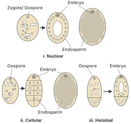
Drawn by us to help you understand the concept clearly, and verified to make sure it's accurate. For exams, practice from your textbook's own diagram.
What this figure shows. This diagram compares the three types of endosperm development, shown side by side as nuclear, cellular, and helobial, each traced from the oospore (zygote) stage through to a distinct pattern of endosperm and embryo formation, the nuclear type showing free nuclei before wall formation, the cellular type showing walls forming from the first division onward, and the helobial type …
Mosaic Endosperm …