Question 76 of 99
Q.Draw and label the structure of Embryo sac.
Puducherry TnboardTamil Nadu HSC (DGE) Board 2022Subjective· 3mImportance★★★★★
77% · 76/99 Questions
You're viewing a preview — the full solution, concept, methods & PYQ mapping are locked.
Start your 14-day free trial to unlock the full solution →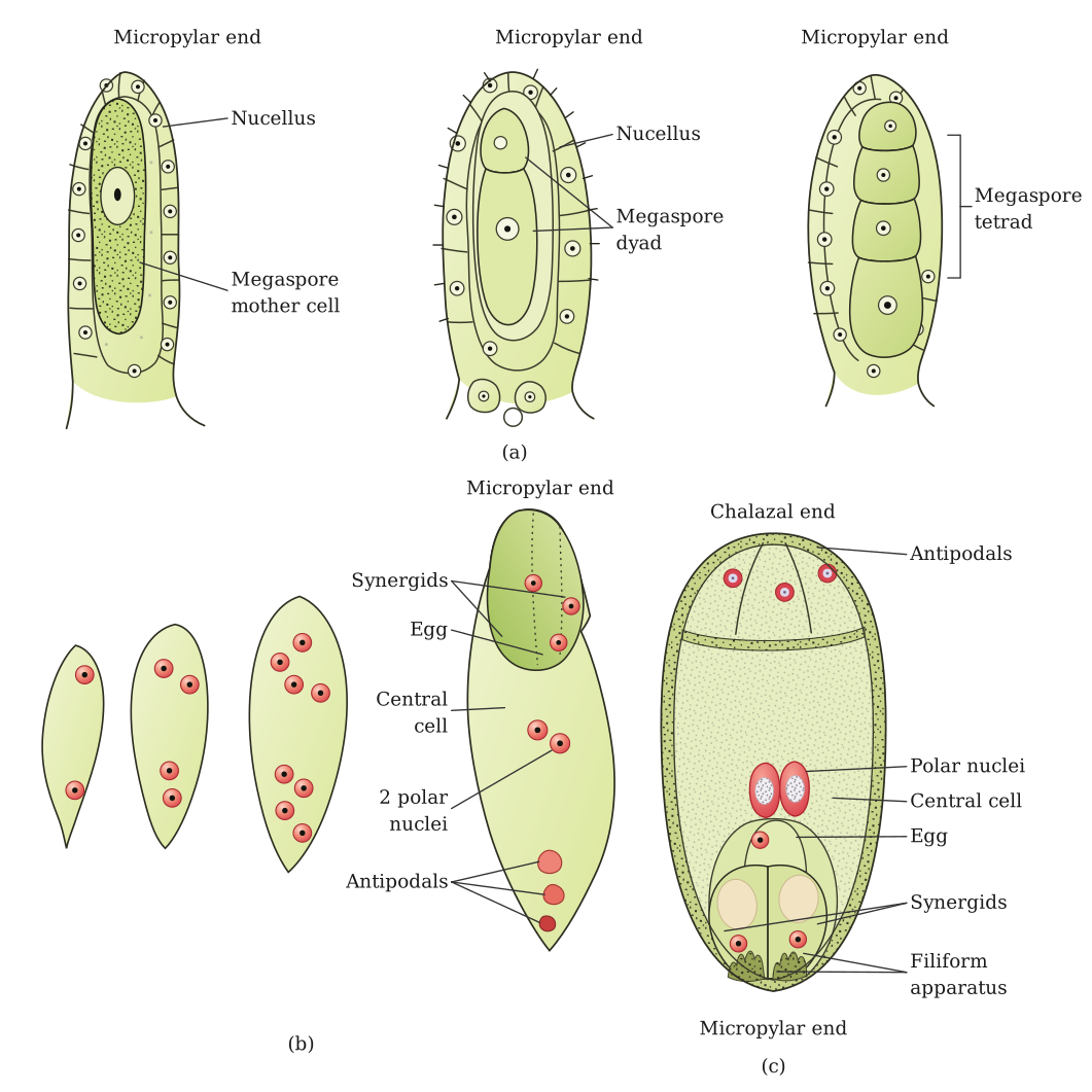
The mature angiosperm embryo sac is a 7-celled, 8-nucleate structure with an egg apparatus, antipodals and a central cell bearing two polar nuclei.
Since a diagram cannot be physically drawn here, the structure of the mature (Polygonum-type) embryo sac is described part by part, exactly as it would be labelled in a diagram:
- Egg apparatus (micropylar end): situated at the end of the embryo sac nearest the micropyle, this group of three cells consists of one central egg cell (oosphere) flanked on either side by two synergid cells. The synergids possess a specialised thickened structure called the filiform apparatus at their micropylar tip, which helps guide the entry of the pollen tube into the embryo sac during fertilisation.
- Antipodal cells (chalazal end): at the opposite end of the embryo sac, nearest the chalaza, lie three antipodal cells, which are thought to have a nutritive role in the developing embryo sac but usually degenerate before or soon after fertilisation. …
Unlock everything free for 14 days
- Full step-by-step solutions
- Concept-first explanations
- Methods, shortcuts & mistakes
- PYQ mapping + timed mock tests
Full access for 14 days. No credit card required.