Biology · Ch 9 — Control and Co-ordination
Pituitary Gland: Structure and Portal System
Pituitary Gland: Structure and Portal System
B. Pituitary gland or hypophysis gland : The pituitary gland (hypophysis) is the smallest endocrine gland, a pea-sized, reddish-grey structure that controls almost every other endocrine gland -- earning it the old nickname 'master endocrine gland' -- lying just below the hypothalamus, to which it is attached by the infundibulum (hypophyseal stalk), and lodged in a bony depression of the sphenoid bone called the sella turcica.
The gland has two lobes of entirely different embryonic origin -- a dual origin. The adenohypophysis (anterior lobe) develops as an upward-growing outpocketing of the buccal-cavity roof called Rathke's pouch, which loses its original connection to the mouth as the embryo develops; it is a highly cellular, vascular tissue containing acidophil, basophil and chromatophore secretory cells, and is itself subdivided into pars distalis, pars intermedia and pars tuberalis. The neurohypophysis (posterior lobe) instead develops as a downward-growing extension of the hypothalamus, and stays connected to it directly by axon fibres; it is further divided into the pars nervosa (neural lobe), the infundibulum and the median eminence. A small, poorly-developed intermediate lobe (pars intermedia) sits in the cleft between the two main lobes.
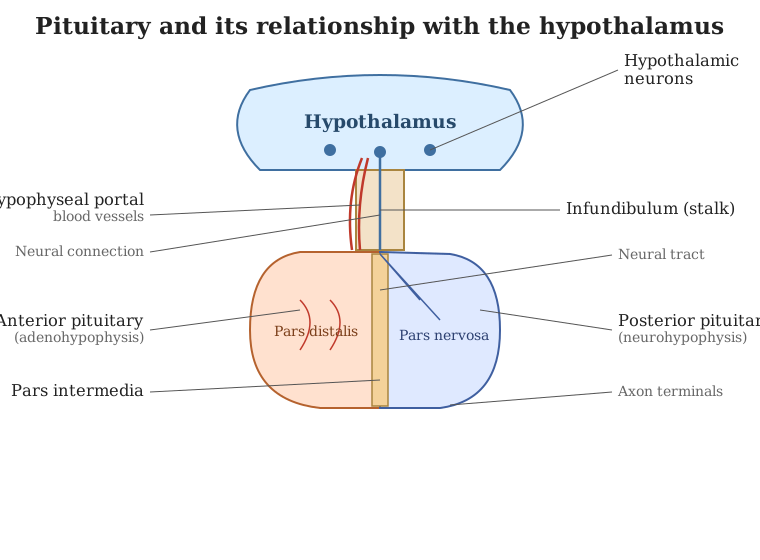
Drawn by us to help you understand the concept clearly, and verified to make sure it's accurate. For exams, practice from your textbook's own diagram.
What this figure shows. A midline diagram of the pituitary gland hanging from the hypothalamus by the infundibulum, showing the optic chiasma just above it; the supraoptic and paraventricular nuclei of the hypothalamus (which make ADH and oxytocin respectively) sending axons down through the median eminence to the pars nervosa of the neurohypophysis, from where they are released into the hypophyseal vein; and, alongside, the hypophyseal portal vein carrying releasing/inhibiting hormones down from the hypothalamus to the pars distalis of the adenohypophysis, with the small pars intermedia shown between the two lobes and the hypophyseal artery supplying the whole gland.
9.25: Pituitary gland. …
…