Biology · Ch 9 — Control and Co-ordination
Thyroid Gland: Structure and Hormones
Thyroid Gland: Structure and Hormones
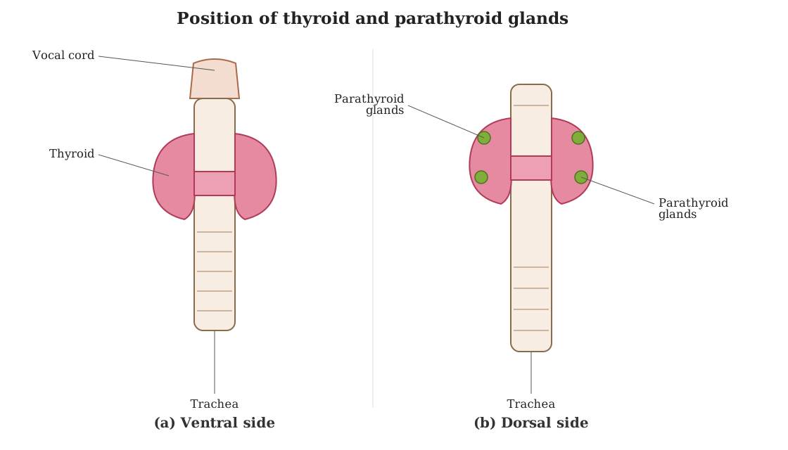
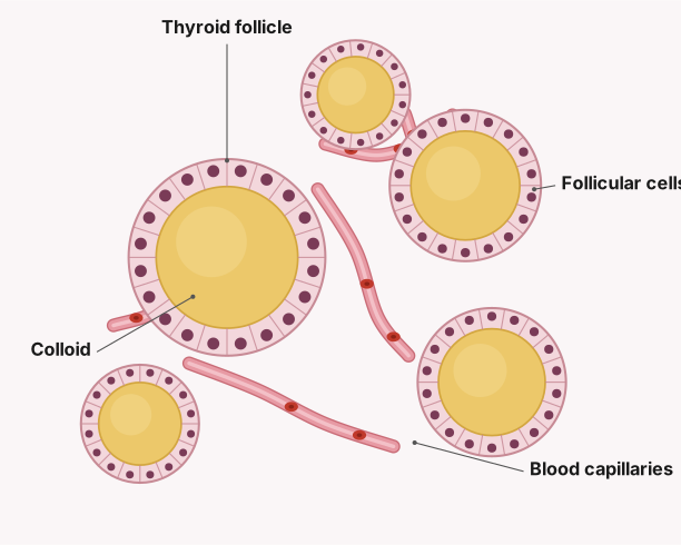
D. Thyroid gland : The thyroid gland is the largest endocrine gland, weighing about 25 grams, bilobed and richly vascular, sitting in front of the trachea just below the larynx, its two lobes joined by a non-secretory band, the isthmus. The lobes are made of rounded follicles held together by connective-tissue stroma (which contains blood capillaries and scattered parafollicular, or 'C', cells); each follicle is a single layer of cuboidal epithelium resting on a basement membrane, enclosing a lumen filled with hormone-rich colloid.
Drawn by us to help you understand the concept clearly, and verified to make sure it's accurate. For exams, practice from your textbook's own diagram.
What this figure shows. Two views of the thyroid gland: (A) its gross external anatomy in the neck, showing the left and right thyroid lobes lying against the trachea just below the larynx, joined across the front of the trachea by a narrow isthmus; and (B) a magnified histological view of thyroid tissue showing rounded thyroid follicles, each a ring of cuboidal follicular cells enclosing a colloid-filled lumen, surrounded by a network of blood capillaries and scattered parafollicular ('C') cells lying between the follicles.
9.28: Thyroid gland. …
Drawn by us to help you understand the concept clearly, and verified to make sure it's accurate. For exams, practice from your textbook's own diagram.
What this figure shows. Fig. 9.28 B: the thyroid's rounded follicles, each a single layer of cuboidal follicular cells around a lumen filled with hormone-rich colloid, with blood capillaries in the connective-tissue stroma between the follicles. …
Stimulated to secrete by thyroid stimulating hormone (TSH) from the pituitary, the follicular cells make two hormones: thyroxine (tetraiodothyronine/T4, carrying four iodine atoms) and triiodothyronine (T3, carrying three). Both are made by enzymatically attaching iodine atoms to the amino acid tyrosine -- one iodine gives monoiodothyronine (T1), two give diiodothyronine (T2), and T1/T2 molecules then join end to end and are further metabolised inside the follicle's colloidal mass to yield T3 and T4; T3, though secreted in smaller quantity, is physiologically the more active of the two. Uniquely among endocrine glands, the thyroid actually stores its hormones (as colloid) before secreting them, and their release is governed by pituitary thyrotropin through negative feedback. …