Biology · Ch 2 — Reproduction in Lower and Higher Animals
Structure of the Secondary Oocyte
Structure of the Secondary Oocyte
Structure of secondary oocyte : In humans, what is popularly called the 'unfertilized egg' — the cell released from the ovary at ovulation — is, strictly, the secondary oocyte. It is non-cleidoic (without a shell) and microlecithal (containing only a very small quantity of yolk), roughly 0.1 mm (100 microns) in size. It is rounded, non-motile, haploid and devoid of centrioles. Its nucleus appears unusually large and is called the germinal vesicle; a typical nucleus (pronucleus) is only formed at the time of fertilization. The cytoplasm of the egg is called the ooplasm.
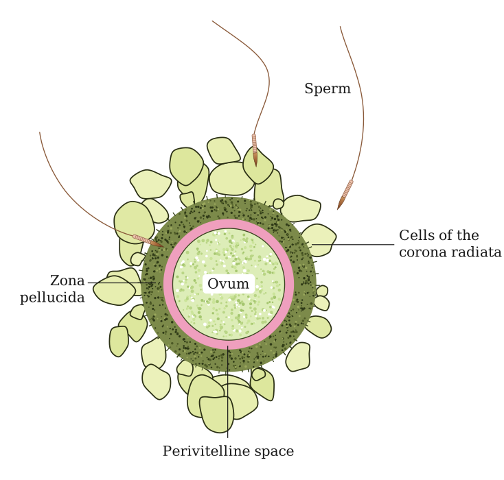
The egg is surrounded by several coverings. Immediately around the cell membrane, the vitelline membrane secretes a non-cellular, glycoprotein-rich membrane on its outside, the zona pellucida. Adhering to the outer surface of the zona pellucida are several radially elongated cells, together forming the corona radiata — these cells are derived from the innermost layer of granulosa cells of the follicle, and are held firmly to the zona pellucida and to each other by hyaluronic acid (a mucopolysaccharide). Between the vitelline membrane and the zona pellucida lies a fluid-filled space, the perivitelline space, in which the first polar body lies. …
Drawn by us to help you understand the concept clearly, and verified to make sure it's accurate. For exams, practice from your textbook's own diagram.
What this figure shows. The ovulated secondary oocyte with its coverings in the order the text describes them: the radially elongated cells of the corona radiata outside, the non-cellular zona pellucida, and the perivitelline space between it and the egg membrane, with sperms approaching. The textbook's Fig. 2.12 draws the same egg without sperms and also marks the germinal vesicle, the vitell …