Biology · Ch 1 — Reproduction in Lower and Higher Plants
Androecium and Structure of Anther
Androecium and Structure of Anther
The androecium is the male reproductive whorl of the flower; each individual member of this whorl is called a stamen. A typical stamen has three parts, a slender filament, a connecting tissue called the connective, and, at its tip, the anther, which bears two lobes (thecae).
A mature anther is usually described as dithecous (two-lobed) and tetrasporangiate, meaning each of its two lobes contains two pollen sacs, giving four pollen sacs in total across the whole anther. In its earliest, immature state, the anther is simply a compact mass of parenchyma cells enclosed by a single layer of protective epidermis. Differentiation begins when certain cells just beneath the epidermis (hypodermal cells) transform into archesporial cells, setting up the internal specialisation that follows.
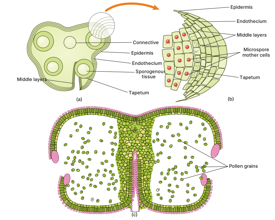
T. S. of Anther : Each archesporial cell then divides to produce an inner sporogenous cell and an outer primary parietal cell. The sporogenous cell proliferates into sporogenous tissue, and every cell of this tissue is capable of eventually giving rise to a tetrad of microspores. The primary parietal cell, meanwhile, divides repeatedly to build up the anther wall, which in a mature anther comprises four distinct layers, from outside in:
- the epidermis, the outermost layer of flattened, protective cells;
- the endothecium, a sub-epidermal layer of radially elongated cells with fibrous wall thickenings that help the anther split open (dehisce) at maturity;
- the middle layers, one or two layers of thin-walled cells lying just inside the endothecium, which often disintegrate as the anther matures; and
- the tapetum, the innermost, nutritive layer that directly surrounds and nourishes the developing sporogenous tissue (the microspore mother cells) as they undergo meiosis.
Drawn by us to help you understand the concept clearly, and verified to make sure it's accurate. For exams, practice from your textbook's own diagram.
What this figure shows. Panel (a) is a transverse section of a young, dithecous anther: the four pollen sacs (microsporangia) grouped in two lobes around the central connective, each sac bounded from outside in by the epidermis, the endothecium, the middle layers and the innermost nutritive tapetum, which directly encloses the sporogenous tissue. Panel (b) magnifies one microsporangium wall so the four wall layers and the microspore mother cells inside can be told apart. Panel (c) is the mature, dehisced anther after the endothecium has dried and each lobe has split open along the line between its two pollen sacs, with the pollen grains spilling out. Panels (a) and (c) carry exactly the content of the textbook's Fig. 1.4 (a) 'T. S. of anther' and Fig. 1.4 (b) …
…