Chemistry · Ch 14 — Biomolecules
Secondary structure of proteins
Secondary structure of proteins
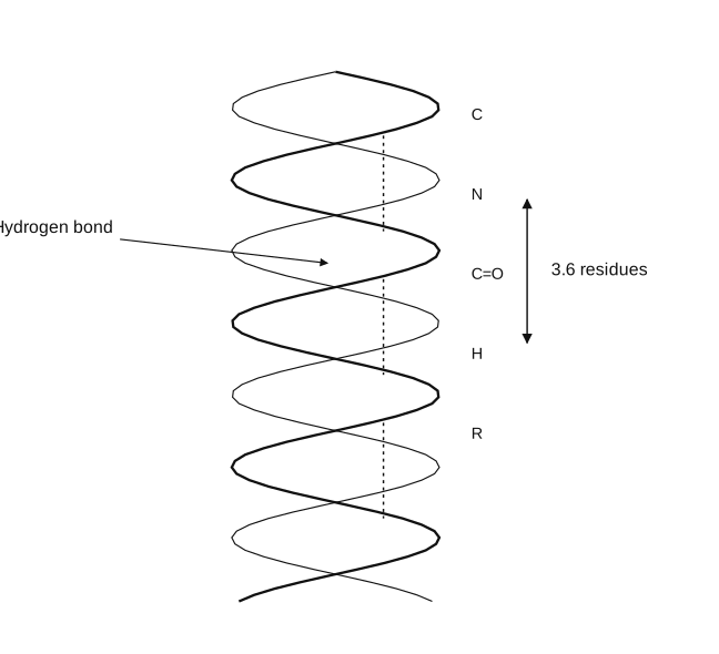
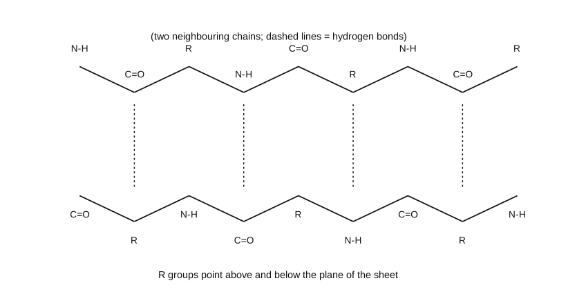
The secondary structure of a protein describes the regular, LOCAL three-dimensional shape that a stretch of the polypeptide backbone folds into, distinct from (and nested within) the larger overall tertiary fold of the whole chain. It arises specifically from hydrogen bonding between the N-H proton of one peptide (amide) linkage and the C=O oxygen of a DIFFERENT peptide linkage elsewhere along the same backbone (or, for the beta-sheet, along a neighbouring backbone) -- crucially, this is backbone-to-backbone hydrogen bonding, not a side-chain interaction, which is why secondary structure is largely independent of which particular amino acids (i.e. which R groups) are actually present. Two secondary-structure patterns recur throughout the protein world. The alpha-helix forms when a single polypeptide chain twists into a right-handed (clockwise) spiral; each complete turn of this spiral contains 3.6 amino-acid residues, and the defining hydrogen-bonding pattern links the C=O group of one residue to the N-H group of the residue FOUR positions further along the same chain -- these hydrogen bonds run roughly parallel to the helix's long axis, holding the coil rigid, while the bulkier R (side-chain) groups all point outward, away from the helix's core. Myosin (in muscle) and alpha-keratin (in hair) are proteins built almost entirely of alpha-helical secondary structure. The beta-pleated sheet forms instead when two or more separate polypeptide strands (which may be different regions of one long chain folded back on itself, or genuinely separate chains) line up SIDE BY SIDE; here the C=O and N-H bonds of the backbone lie roughly IN the plane of the resulting sheet, and hydrogen bonding occurs ACROSS between neighbouring strands (an N-H on one strand to a C=O on the next, and so on), while the R groups on each strand project alternately above and below the sheet's plane. Because bulky side chains would clash with this side-by-side packing, the beta-pleated arrangement is specifically favoured at stretches of chain where the amino acids present have small R groups. Real proteins very often contain regions of BOTH alpha-helix and beta-sheet, plus additional 'random' regions that fit neither pattern -- spider dragline silk protein is a striking example, combining beta-pleated-sheet regions (which give it high tensile strength) with alpha-helical regions …
Drawn by us to help you understand the concept clearly, and verified to make sure it's accurate. For exams, practice from your textbook's own diagram.
What this figure shows. A schematic of the polypeptide backbone coiled into a right-handed spiral (the alpha-helix), with R groups labelled projecting outward from the coil at regular intervals and H atoms shown along the backbone. A bracket marks '3.6 residues' as the length of one complete turn of the helix, and a dashed line labelled 'Hydrogen bond' runs roughly parallel to the helix's long axis, connecting a C=O group on one turn to an N-H group one turn further along (i.e. four residues along the chain), illustrating exactly th …
Drawn by us to help you understand the concept clearly, and verified to make sure it's accurate. For exams, practice from your textbook's own diagram.
What this figure shows. Two (or more) extended, zig-zag polypeptide strands drawn side by side and roughly parallel, each strand's backbone alternating N-H and C=O groups (with R groups, not individually drawn in detail here, understood to project alternately above and below the sheet's plane). Dashed lines mark hydrogen bonds running ACROSS between the strands -- from an N-H on one strand to a C=O on the neighbouring strand, and vice versa at the next position along -- the inter-strand H-bonding pattern that holds the pleated-sheet arrangement together, distinct fro …
Worked out. In collagen, the structural protein of connective tissue, each individual polypeptide chain first coils into an unusual LEFT-handed helix (unlike the right-handed alpha-helix of most other proteins); then three of these left-handed chains wind around one another into an overall RIGHT-handed triple helix. This nested left-then-right helical architecture is unique to collagen among the secondary/tertiary structures described in this section and gives collagen fibres their characteristi …