Chemistry · Ch 14 — Biomolecules
Tertiary structure of proteins
Tertiary structure of proteins
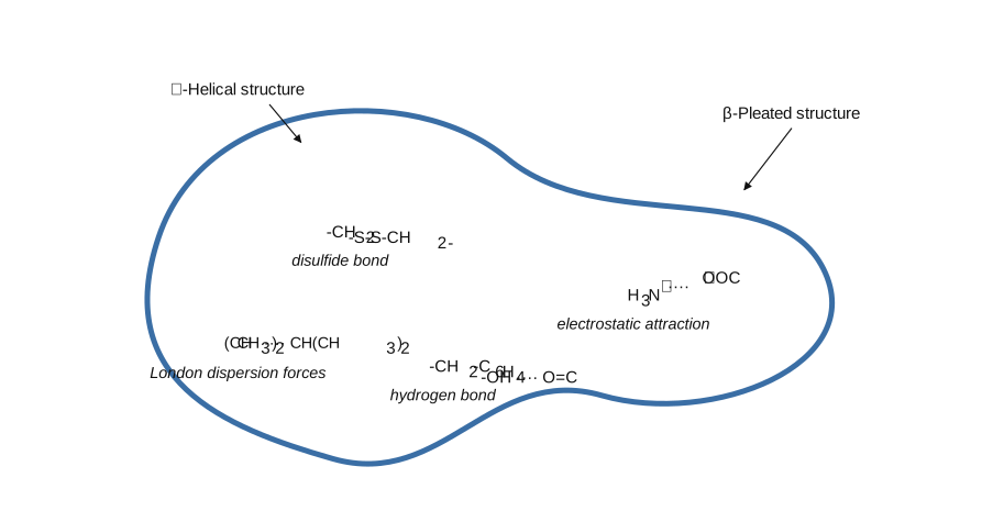
The tertiary structure of a protein is the overall, non-repeating three-dimensional shape adopted by one ENTIRE polypeptide chain -- the result of the whole chain folding up in a specific way that both stabilises the folded structure itself and allows it to interact favourably with the surrounding aqueous (water-based) environment of the cell. It is precisely this tertiary-level folding that produces the two broad molecular shape-classes introduced back in section 14.3.3: a chain that folds compactly into a sphere gives a globular protein, while chains held in an extended, parallel arrangement give a fibrous protein. Several distinct kinds of force, acting between side chains (R groups) that may be far apart in the primary sequence but end up brought close together once the chain is folded, cooperate to stabilise a particular tertiary structure: hydrogen bonding (between polar side-chain groups); dipole-dipole attraction (arising from polar bonds within side chains); electrostatic attraction (between oppositely-charged ionic side-chain groups, such as a -COO- and an -NH3+ brought into proximity); and London dispersion forces (weak, universal attractions between any nearby non-polar side chains). Uniquely among these, one further stabilising force is a genuine COVALENT bond rather than a weaker non-covalent interaction: the disulfide bond, formed when two -SH (thiol) groups on two nearby cysteine residues are oxidatively coupled together into an -S-S- linkage -- because it is a true covalent bond, a disulfide b …
Drawn by us to help you understand the concept clearly, and verified to make sure it's accurate. For exams, practice from your textbook's own diagram.
What this figure shows. A schematic globular protein chain folded into a compact 3-D shape, with regions of alpha-helical structure and beta-pleated structure both visible as sub-regions of the same folded chain (illustrating that a single tertiary structure typically incorporates both kinds of secondary structure). Labelled arrows point to four different stabilising-force examples found at different points in the fold: a disulfide bond (a direct covalent S-S link between two cysteine side chains), an electrostatic attraction (between an ionic side-chain pair), London dispersion forces (between nearby non-polar side chains) and a hydrogen bond (between polar groups) -- …