Biology · Ch 10 — Human Health and Diseases
Filariasis (Elephantiasis)
Filariasis (Elephantiasis)
D. Filariasis / Elephantiasis : Filariasis, also called elephantiasis in its classic form, is caused by thread-like nematode worms transmitted from person to person by mosquito bite. It has three recognised subtypes — lymphatic filariasis, subcutaneous filariasis (e.g. caused by Loa loa, Mansonella species) and serous (abdominal) cavity filariasis (e.g. Mansonella species) — of which lymphatic filariasis, caused by Wuchereria bancrofti, Brugia malayi and Brugia timori, is the classic 'elephantiasis'.
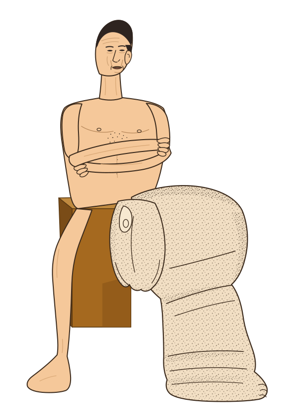
Signs and symptoms: Oedema with thickening of the skin and underlying tissue; W. bancrofti particularly affects the legs, arms, breasts and scrotum. Worms infecting the lymphatic system cause enlargement of lymph vessels and nodes, and the resulting swelling can make limbs resemble an elephant's legs — the classic elephantiasis. Accumulation of lymph fluid in tissue (lymphoedema) causes further swelling, and fluid accumulation around the testis causes hydrocele (enlarged testes).
Drawn by us to help you understand the concept clearly, and verified to make sure it's accurate. For exams, practice from your textbook's own diagram.
What this figure shows. The swollen, thickened lower limb of lymphatic filariasis - the 'elephant leg' that gives elephantiasis its name - caused by adult worms blocking the lymph vessels so that lymph acc …
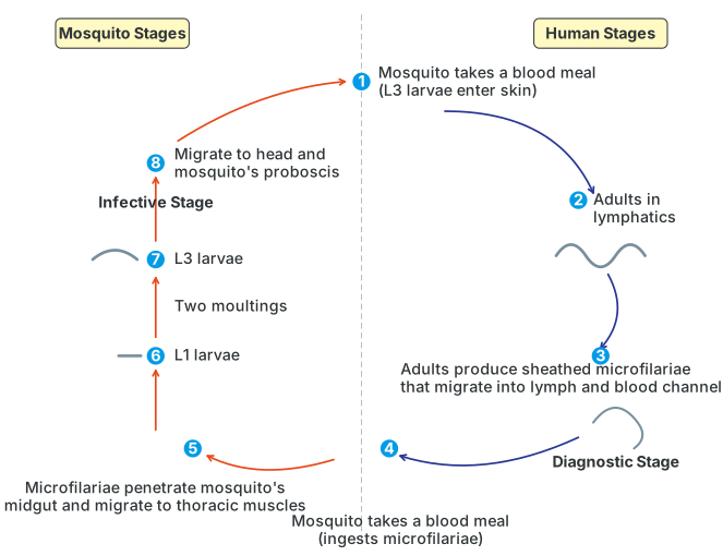
Mode of transmission: W. bancrofti is transmitted to humans by the female Culex mosquito. Infective larvae escape from the mosquito onto human skin, penetrate it, undergo two moultings as they develop into adults, and settle in the lymphatic system; the incubation period can be as long as 8-16 months.
Drawn by us to help you understand the concept clearly, and verified to make sure it's accurate. For exams, practice from your textbook's own diagram.
What this figure shows. A cyclic diagram, numbered through eight stages, split into 'Mosquito Stages' and 'Human Stages'. It shows a mosquito taking a blood meal from an infected human and ingesting microfilariae (the diagnostic stage circulating in that person's blood/lymph); inside the mosquito, these microfilariae penetrate the midgut and migrate to the thoracic muscles, developing through an L1 larval stage, two moultings, and an infective L3 larval stage that migrates to the mosquito's head and proboscis. The cycle then shows the same mosquito taking a blood meal from a new, healthy person, during which the L3 larvae enter the skin (the infective stage); these larvae are traced developing into adult worms that settle in the lymphatics, where the adults produce new sheathed microfilariae that migrate into the lymph and …
…