Biology · Ch 3 — Inheritance and Variation
Chromosomes
Chromosomes
Can you recall?
- What is a chromosome?
- How many chromosomes are present in a human somatic cell and in a reproductive cell?
Chromosomes are thread-like (filamentous) bodies found in the nucleus of eukaryotic cells; the name itself (Greek chromo = colour, soma = body) was coined by W. Waldeyer in 1888 because these bodies stain readily with certain dyes. They vary in size across species (roughly 0.1 to 33 µm in length and 0.2 to 2 µm in thickness at metaphase), are visible under the microscope specifically during cell division (being most condensed and clearly countable at metaphase), and are chemically built from DNA together with histone and non-histone proteins. Beyond simply carrying hereditary information, chromosomes are self-replicating and are central to heredity, mutation, variation and the evolutionary development of eukaryotic species.
Function : Chromosomes mainly act as the carriers of heredity.
Number of chromosomes : The number of chromosomes of a species is constant and specific, which is why chromosome counts are useful in studying phylogeny and taxonomy. 'Ploidy' describes how many times the basic haploid chromosome set (x) is repeated in a cell. When the chromosome number is an exact multiple of x, the cell is euploid — covering monoploid/haploid (n, one set), diploid (2n, two sets), triploid (3n), tetraploid (4n) and so on. When the number departs from an exact multiple of the haploid set by the gain or loss of one or more individual chromosomes, the cell is aneuploid — further split into hyperploid conditions such as trisomy (2n+1) and tetrasomy (2n+2), and hypoploid conditions such as monosomy (2n−1) and nullisomy (2n−2). (See Chart 3.5.)
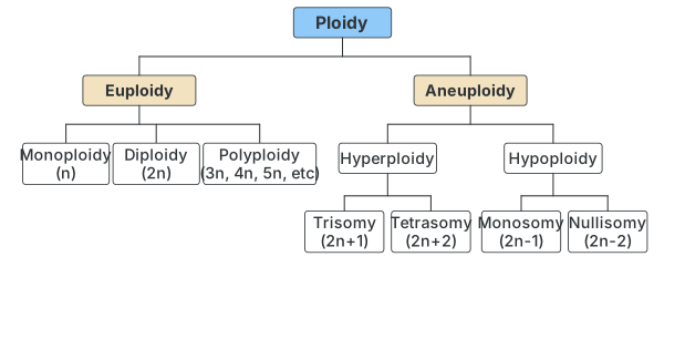
Chart 3.5 : Variation in chromosome number (ploidy). Ploidy branches into Euploidy (Monoploidy n, Diploidy 2n, Polyploidy 3n/4n/5n...) and Aneuploidy, which branches into Hyperploidy (Trisomy 2n+1, Tetrasomy 2n+2) and Hyp …
Always Remember
- Genes and chromosomes always occur in pairs in a diploid organism.
- Alleles located on a chromosome segregate along with the chromosome during gamete formation.
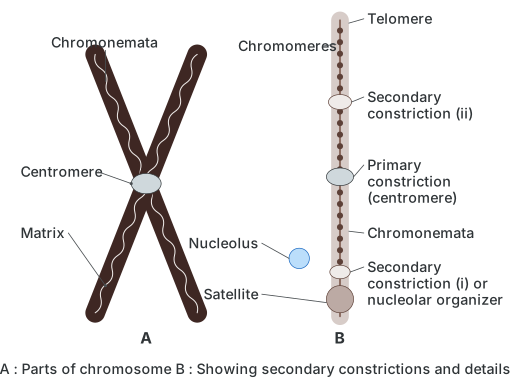
Structure of chromosome : Chromosomes are best visible under the microscope at metaphase, when they are most condensed. A typical metaphase chromosome consists of two chromatids joined at a primary constriction, the centromere, whose disc-shaped kinetochore is where spindle fibres attach during cell division. Some chromosomes carry one or two additional secondary constrictions: at secondary constriction I the nucleolus reorganises during interphase, while a small satellite body (SAT body) is occasionally found attached beyond secondary constriction II. Each chromatid itself contains a long, coiled, unbranched DNA thread called the chromonema running its full length, made of a double-stranded DNA molecule stretching end to end; the free ends of the chromosome are called telomeres.
Depending on where the centromere sits, chromosomes are classified into four shapes: metacentric (centromere central, V-shaped), submetacentric (centromere off-centre, L-shaped), acrocentric (centromere near one end, J-shaped) and telocentric (centromere at the very tip, I-shaped, or rod-like).
Drawn by us to help you understand the concept clearly, and verified to make sure it's accurate. For exams, practice from your textbook's own diagram.
What this figure shows. Diagrams a metaphase chromosome made of two sister chromatids joined at the centromere (primary constriction), each chromatid carrying a long coiled DNA thread called the chromonema studded with chromomeres, with the free ends called telomeres, and marks the secondary constriction where the nucleolar organiser sits and, in a few chromosomes, a satellite body attached beyond a second secondary constriction. …
Activity
Study the types of chromosome according to the position of the centromere. Observe and complete the table with three columns: type of chromosome (sketch), name of chromosome, position of centromere. Rows to complete: metacentric (centromere in the middle), submetacentric (slightly away from the middle), acrocentric (near one end), telocentric (at one end). …
Drawn by us to help you understand the concept clearly, and verified to make sure it's accurate. For exams, practice from your textbook's own diagram.
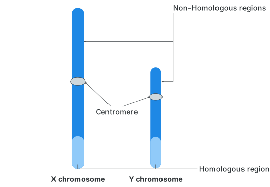
What this figure shows. Compares the human sex chromosomes side by side: the X is a longer, straight, metacentric chromosome with mostly active euchromatin, while the Y is shorter and acrocentric with mostly inactive heterochromatin; both share a small homologous region (where crossing over can occur) alongside their much larger non-homologous regions that carry their own …