Biology · Ch 6 — Plant Water Relation
Structure of Stomatal Apparatus
Structure of Stomatal Apparatus
Do you know?
- The number of stomata per unit area of leaf is called stomatal frequency.
- The correlation between the number of stomata and the number of epidermal cells per unit area is called stomatal index (I).
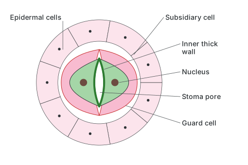
A typical stomatal apparatus in the leaf epidermis is built from three components: a pair of guard cells, the stoma (the pore they enclose between them), and a set of surrounding accessory (or subsidiary) cells.
Drawn by us to help you understand the concept clearly, and verified to make sure it's accurate. For exams, practice from your textbook's own diagram.
What this figure shows. This close-up diagram of a single guard cell, set among its neighbouring epidermal and subsidiary cells, labels the nucleus, the guard cell's characteristically uneven wall (marked as an inner thick wall bordering the stomatal pore), and the pore itself. It is used to show, at the level of one cell, exactly which wall of the guard cell is reinforced and which is thin, since that asymmetry is what allows the cell to bow outw …
Drawn by us to help you understand the concept clearly, and verified to make sure it's accurate. For exams, practice from your textbook's own diagram.
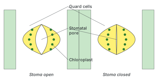
What this figure shows. This is a paired before-and-after diagram showing the same pair of guard cells in two states: turgid, where their thin outer walls bulge outward and their thickened inner walls are pulled apart to leave a visibly open elliptical pore between them, and flaccid, where the guard cells relax back and the pore narrows almost completely shut. Placing the two states side by side is how the figure demonstrates that stomatal opening and closing is purely a matter of the guard cells' …
Guard cells are living, nucleated cells, distinguished from ordinary epidermal cells by having an unevenly thickened wall: the inner wall, the one facing directly onto the stomatal pore, is comparatively thick and inelastic, while the outer, lateral wall (facing away from the pore, toward the neighbouring epidermal cells) is comparatively thin and elastic. Guard cells also contain a small number of chloroplasts, capable of only limited photosynthesis, which is unusual since most epidermal cells lack chloroplasts altogether. This combination of an uneven wall and the ability to change shape is what allows guard cells to open and close the pore between them: as internal turgor rises or falls, the thin elastic outer wall stretches or relaxes, and because of the guard cell's characteristic curved shape, this pulls the thick inner walls apart to widen the pore, or lets them come back together to narrow it.
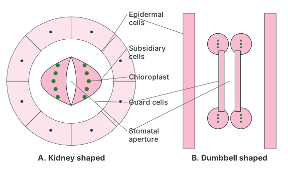
Guard-cell shape differs between major plant groups: in dicotyledons, guard cells are typically kidney- (bean-) shaped, enclosing an elliptical pore between them, while in grasses and other monocotyledons, guard cells are typically dumbbell- (barbell-) shaped, with bulbous ends and a narrower waist in the middle. Some plants, such as Cyperus, are unusual in possessing both kidney-shaped and dumbbell-shaped guard cells within the same leaf. In Cyperus, both kidney- and dumbbell-shaped guard cells are present.
Drawn by us to help you understand the concept clearly, and verified to make sure it's accurate. For exams, practice from your textbook's own diagram.
What this figure shows. This figure compares two guard-cell shapes side by side, each shown with its flanking epidermal and subsidiary cells and internal chloroplasts. One pair is drawn as classic kidney- (bean-) shaped guard cells enclosing an elliptical stomatal aperture, typical of dicotyledonous plants, while the other pair is drawn as dumbbell- (barbell-) shaped guard cells with bulbous ends and a narrow waist, typical of grasses and other monocotyledons. The side-by-side layout is what lets a student see directly how the same functional job — opening and closing a pore by changes in turgor — is carried out by two structurally diff …
…