Q.Explain the histological structure of testis.
Concept understanding — Male Reproductive System
The male reproductive system centres on the paired testes, each divided by internal septa into 200-250 lobules containing seminiferous tubules, the site of sperm production, which alone make up about 80% of testicular tissue. The tubule wall is lined by Sertoli (nurse) cells, which nourish developing sperm and secrete inhibin (a negative-feedback hormone that regulates sperm output), while Leydig (interstitial) cells sitting between the tubules secrete testosterone, the androgen that drives spermatogenesis. The testes sit outside the abdominal cavity in the scrotum, a skin sac that keeps them 2-3 degrees C cooler than internal body temperature — a genuine requirement, since viable sperm cannot form at normal body heat; a testis that fails to descend (cryptorchism) can cause sterility if not corrected early. Sperm travel from the tubules through the rete testis and vasa efferentia into the epididymis, where they are stored and physiologically mature, then on through the vas deferens and ejaculatory duct to the urethra. Semen is formed by adding the secretions of three accessory glands to the sperm: the paired seminal vesicles (the largest fluid contributor, supplying fructose, ascorbic acid, prostaglandins and a coagulating enzyme), the single prostate gland (a slightly acidic, citrate- and enzyme-rich secretion), and the paired bulbourethral glands (lubrication). The penis, the copulatory organ, has an enlarged glans covered by the foreskin.
The testis is wrapped in three coverings (tunica vaginalis, albuginea, vasculosa) and internally divided into lobules of coiled seminiferous tubules (germinal epithelium + Sertoli cells) with hormone-secreting Leydig cells between them.
Outer tunica vaginalis → tunica albuginea → tunica vasculosa internally; septa create 200–300 lobules with coiled seminiferous tubules (germinal epithelium + Sertoli cells); interstitial (Leydig) cells lie between tubules, secreting testosterone.
The testis is wrapped in three coverings (tunica vaginalis, albuginea, vasculosa) and internally divided into lobules of coiled seminiferous tubules (germinal epithelium + Sertoli cells) with hormone-secreting Leydig...
Step 1. Externally, the testis is covered by three layers: the tunica vaginalis, an incomplete peritoneal covering; the tunica albuginea, a tough collagenous connective-tissue layer beneath it; and, innermost, the tunica vasculosa, a thin, membranous and vascular layer.
Step 2. Fibrous septa arising from the tunica albuginea divide the testis internally into about 200–300 testicular lobules, each containing 1–4 highly coiled seminiferous tubules.
Step 3. Each seminiferous tubule is lined internally by cuboidal germinal epithelial cells (spermatogonia), interspersed with fewer, larger, pyramidal Sertoli (sustentacular) cells that nourish the developing sperm; the successive stages of spermatogenesis are visible within the tubule wall, from spermatogonia near the base to maturing sperm near the lumen.
Step 4. In the loose connective tissue between the seminiferous tubules lie the interstitial or Leydig cells, which secrete the male hormone testosterone.
The testis's histology, from outside in, is tunica vaginalis → tunica albuginea → tunica vasculosa, with the albuginea's septa dividing the interior into lobules of coiled, germinal-epithelium-and-Sertoli-cell-lined seminiferous tubules, and testosterone-secreting Leydig cells filling the spaces between tubules.
Describe the coverings, then the internal lobular/tubular structure, then the interstitial cells
- Mixing up the order of the three coverings (vaginalis outermost, albuginea middle, vasculosa innermost).
- Placing the Leydig (interstitial) cells inside the seminiferous tubule instead of in the connective tissue between tubules.
- CBSE 2026Set ANNUAL1 markQ.Give the function of Leydig's cells.
›Reveal solutionSolution
Leydig's cells (interstitial cells), located in the interstitial spaces between the seminiferous tubules of the testis, secrete the male sex hormone testosterone, which is essential for spermatogenesis and the develop...
Leydig's cells are the testosterone-secreting endocrine cells of the testis.
Leydig cells (also called interstitial cells) are found scattered in the loose connective tissue occupying the spaces between the seminiferous tubules within the testicular lobules. Under the stimulation of luteinizing hormone (LH, also called interstitial cell stimulating hormone or ICSH in males) from the anterior pituitary, Leydig cells synthesize and secrete testosterone, the principal male androgen. Testosterone regulates spermatogenesis (in conjunction with FSH acting on Sertoli cells), promotes the growth and function of male accessory reproductive organs, and is responsible for the development and maintenance of male secondary sexual characteristics such as facial/body hair, deepening of voice and muscular development.
✓Final answerLeydig's cells (interstitial cells), located in the interstitial spaces between the seminiferous tubules of the testis, secrete the male sex hormone testosterone, which is essential for spermatogenesis and the development/maintenance of male secondary sexual characters.
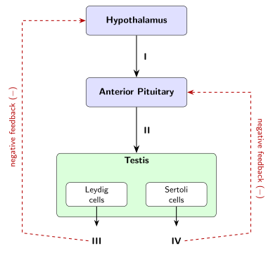
- CBSE 2026Set SEM31 markMCQQ.State the names of the hormones I, II, III, IV according to the following diagram :(a) (I) — Inhibin, (II) — Testosterone, (III) — FSH, (IV) — GnRH(b) (I) — Testosterone, (II) — Inhibin, (III) — FSH, (IV) — GnRH(c) (I) — GnRH, (II) — Testosterone, (III) — Inhibin, (IV) — FSH(d) (I) — GnRH, (II) — FSH, (III) — Testosterone, (IV) — Inhibin
›Reveal solutionSolution
In the male hormone axis: hypothalamic GnRH → pituitary FSH (and LH) → testis; Leydig cells make testosterone and Sertoli cells make inhibin, which give negative feedback. So I = GnRH, II = FSH, III = Testosterone, IV = Inhibin.
Note: this item carries a figure. A specific figure description was not supplied to me in the batch, so I answer from the standard NCERT male hormonal-control diagram.
The hormonal control of male reproduction (hypothalamo–pituitary–gonadal axis):
- The hypothalamus releases GnRH (Gonadotropin-Releasing Hormone) → labelled I.
- GnRH stimulates the anterior pituitary to secrete the gonadotropins FSH and LH. FSH acts on Sertoli cells → labelled II (FSH).
- LH acts on Leydig (interstitial) cells to secrete Testosterone → labelled III.
- Sertoli cells secrete Inhibin, which suppresses FSH secretion by negative feedback → labelled IV.
Both testosterone and inhibin exert negative feedback on the hypothalamus/pituitary. This ordering matches option (d).
A classic NCERT/CBSE Class 12 Biology diagram-based board question on Human Reproduction.
✓Final answer(d) (I) GnRH, (II) FSH, (III) Testosterone, (IV) Inhibin.
- CBSE 2025Set ANNUAL1 markMCQQ.Part of the sperm containing enzymes for penetration of corona radiata is ______.(a) tail(b) middle part(c) neck(d) head
›Reveal solutionSolution
(d) Head — the acrosome, a cap covering the anterior part of the sperm head, contains hydrolytic enzymes (including hyaluronidase) that digest a path through the corona radiata and zona pellucida of the egg.
The acrosome, located in the sperm head, carries the penetration enzymes.
A mature human sperm has four regions — head, neck, middle piece and tail. The head carries the haploid nucleus and, covering its anterior two-thirds, a membrane-bound cap called the acrosome. The acrosome is essentially a modified lysosome packed with hydrolytic enzymes such as hyaluronidase and acrosin. During fertilisation these enzymes are released (the acrosome reaction) and digest the follicle cells of the corona radiata and the glycoprotein zona pellucida surrounding the ovum, allowing the sperm to penetrate and fuse with the egg's plasma membrane. The middle piece, by contrast, is packed with mitochondria that supply ATP for tail movement, not enzymes.
✓Final answer(d) Head (specifically its acrosomal cap).
- CBSE 2023Set ANNUAL1 markMCQQ.The first ejaculation of the semen is ________.(a) Orchidectomy(b) Infertility(c) Spermarche(d) Azoospermia
›Reveal solutionSolution
Spermarche is the medical term for a male's first ejaculation of semen, occurring at puberty.
At puberty, rising gonadotropin and testosterone levels activate the testes, leading to the onset of spermatogenesis and semen production; the first ejaculation event in this process is termed spermarche, paralleling menarche (the first menstrual period) in girls. Option (a) Orchidectomy is a surgical procedure to remove one or both testes, unrelated to normal puberty. Option (b) Infertility is the inability of a couple to conceive after regular unprotected intercourse, a clinical condition, not a developmental milestone. Option (d) Azoospermia is a pathological condition where semen contains no sperm cells at all, a cause of male infertility, not the definition of first ejaculation.
✓Final answerThe correct option is c) Spermarche.
- CBSE 2023Set ANNUAL1 markMCQQ.The male sex hormone testosterone is secreted from :(a) Epididymis(b) Sertoli cells(c) Prostate gland(d) Leydig cell
›Reveal solutionSolution
Testosterone is secreted by the Leydig (interstitial) cells of the testis.
Within the testis, the seminiferous tubules are surrounded by interstitial tissue containing Leydig cells, which, under stimulation by luteinizing hormone (LH) from the pituitary, synthesise and secrete testosterone — the principal androgen responsible for male secondary sexual characteristics and regulation of spermatogenesis. Option (b) Sertoli cells, located within the seminiferous tubules themselves, provide nutrition and support to developing germ cells and secrete factors like inhibin, but they do not produce testosterone. Option (a) Epididymis is a coiled duct where sperm are stored and undergo maturation, and option (c) Prostate gland is an accessory gland that contributes fluid to semen — neither is an endocrine source of testosterone.
✓Final answerThe correct option is d) Leydig cell.
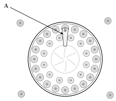
- CBSE 2020Set ANNUAL1 markMCQQ.Identify the cell labelled 'A' in the T.S. of testis (see figure).(a) Leydig cell(b) Basement membrane(c) Sperm(d) Sertoli cell
›Reveal solutionSolution
(d) Sertoli cell — the nurse cell of the seminiferous tubule wall that spans from the basement membrane to the lumen.
A cell within the seminiferous-tubule wall, spanning from the basement membrane to the lumen, is a Sertoli (nurse) cell.
In a T.S. of the testis, each seminiferous tubule wall is lined by germinal epithelium consisting of two main cell types: spermatogenic cells (which divide and mature into sperm, occupying most of the crowded epithelium) and large, tall Sertoli (sustentacular/nurse) cells that rest on the basement membrane and extend right up to the lumen, nourishing and protecting the developing germ cells and secreting fluid that carries sperm. Since the labelled cell 'A' is described as lying at the tubule wall/lumen (i.e., within the tubule's epithelium), it corresponds to a Sertoli cell rather than a Leydig cell, which instead lies outside the tubules in the interstitial connective tissue between them and secretes testosterone.
✓Final answer(d) Sertoli cell — the nurse cell of the seminiferous tubule wall that spans from the basement membrane to the lumen.
- CBSE 2019Set ANNUAL1 markQ.Mention any one function of the Prostate gland.
›Reveal solutionSolution
The prostate gland adds an alkaline secretion to semen that activates the sperm and neutralises vaginal/urethral acidity, aiding sperm survival and motility.
The prostate gland is one of the accessory (secondary) sex glands of the human male reproductive system, surrounding the base of the urethra. Its secretion forms a major part of the seminal plasma.
One function: it secretes a slightly alkaline, milky fluid that (i) forms part of the semen and (ii) neutralises the acidity of the male urethra and the female reproductive tract, thereby activating the sperm and enhancing their motility and survival.
✓Final answerA function of the prostate gland is to secrete an alkaline fluid that forms part of the semen and activates the sperm while neutralising the acidity of the female tract, aiding sperm motility. (Alternative asked: a 'true fruit' is a fruit that develops only from the ripened ovary of the flower after fertilisation, e.g. mango.)
- CBSE 2017Set ANNUAL1 markMCQQ._______ contribute about 60% of the total volume of the semen.(a) Prostate glands(b) Cowper's glands(c) Seminal vesicles(d) Bartholin's glands
›Reveal solutionSolution
(c) Seminal vesicles
Seminal vesicle secretion, rich in fructose, contributes the largest share (about 60%) of semen volume.
Semen is composed of spermatozoa suspended in secretions from several accessory glands. The paired seminal vesicles contribute the largest proportion — roughly 60% of the total semen volume — with a viscous, alkaline, fructose-rich fluid that nourishes sperm and provides energy for their motility. The prostate gland contributes a smaller proportion (roughly 30%) of milky, slightly acidic fluid containing enzymes that help liquefy semen, while the bulbourethral (Cowper's) glands contribute a small amount of clear, lubricating fluid; Bartholin's glands are actually a female reproductive structure and not part of the male semen contribution at all.
✓Final answer(c) Seminal vesicles
- CBSE 2017Set ANNUAL1 markMCQQ.Which part of the sperm produces energy for the movement of sperm ?(a) acrosome(b) nucleus(c) mitochondria(d) axial filament
›Reveal solutionSolution
Mitochondria in the sperm's middle piece supply the energy for its motility.
A human/mammalian sperm's middle piece is densely packed with mitochondria arranged helically around the axial filament (axoneme); these mitochondria carry out oxidative respiration to generate the ATP that powers the beating of the tail (flagellum), propelling the sperm forward. The acrosome contains enzymes for penetrating the egg, the nucleus carries the condensed paternal genetic material, and the axial filament is the contractile microtubule structure that mitochondria-derived ATP actually drives — but it is the mitochondria that are the energy source.
✓Final answerMitochondria — the sperm organelles that generate energy (ATP) for tail movement.
🎓Unlock everything free for 14 days
- ✓Full step-by-step solutions
- ✓Concept-first explanations
- ✓Methods, shortcuts & mistakes
- ✓PYQ mapping + timed mock tests
Full access for 14 days. No credit card required.