Biology · Ch 2 — Reproduction in Lower and Higher Animals
Ovary: Structure and Development
Ovary: Structure and Development
1. Ovary : The ovary is the primary female sex organ, responsible for producing the egg (ovum) and the female reproductive hormones. It is a solid, oval or almond-shaped structure about 3.0 cm long, 1.5 cm broad and 1.0 cm thick, situated in the upper lateral part of the pelvis near the kidney. Each ovary is held in position by ligaments connecting it to the uterus and to the abdominal wall; the largest of these, the broad ligament, is a fold of peritoneum that holds the ovary, oviduct and uterus to the dorsal body wall, while the ovarian ligament specifically attaches the ovary to the uterus. Between them, the two ovaries secrete five hormones — estrogen, progesterone, relaxin, activin and inhibin.
Structure and development of the ovary : Each ovary is differentiated into a central medulla and an outer cortex. The medulla is loose connective tissue (stroma) carrying blood vessels, lymph vessels and nerve fibres, while the cortex, more compact and granular, is packed with numerous tiny masses of cells called ovarian follicles. These arise from immature ova (germ cells) that originate in the dorsal endoderm of the yolk sac during embryonic development and migrate to the gonadal ridge, dividing mitotically along the way; at this stage the cells are called oogonia. As oogonia grow in size, they become surrounded by a layer of granulosa cells, forming the rudiments of the ovarian follicles. This entire process of oogenesis begins well before the birth of the female baby, and by around twelve weeks of foetal development the ovary is fully formed, already containing more than two million primordial follicles.
The cells of the germinal epithelium give rise to groups of oogonia projecting into the cortex as cord-like structures called egg tubes of Pflüger; each cord ends in a round mass of oogonial cells called an egg nest, from which the primordial ovarian follicles develop. Each primordial follicle has, at its centre, a large primary oocyte (2n) surrounded by a single layer of flat follicular cells; this primary oocyte begins meiosis but is arrested at meiosis I. Of the roughly two million primordial follicles present in the foetal ovary, only about one million remain at birth, and only about 60,000–80,000 remain by puberty — the large-scale destruction of primordial follicles during this growth period is called atresia.
Development of primordial follicles into mature Graafian follicles resumes with the onset of puberty. In each menstrual cycle, only one primordial follicle develops into a Graafian follicle, and the two ovaries alternate in producing it from one cycle to the next. The first menstrual cycle, or menarche, normally begins around age 13, and menopause (the stopping of cycles) occurs between ages 45 and 55; the period between menarche and menopause is the female's reproductive age, roughly 32 years, during which she will produce a maximum of about 416 eggs (32 years × 13 cycles per year). …
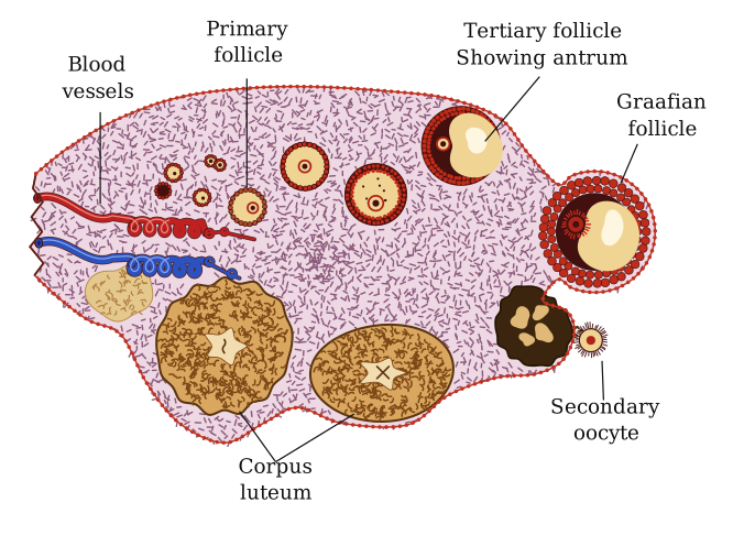
Drawn by us to help you understand the concept clearly, and verified to make sure it's accurate. For exams, practice from your textbook's own diagram.
What this figure shows. A section through the ovary read clockwise like the textbook's Fig. 2.5: primordial and primary follicles in the cortex under the germinal epithelium, a secondary and a tertiary follicle growing an antrum, the mature Graafian follicle bulging at the surface, the ovulated secondary oocyte leaving it, the corpus luteum that forms from the emptied follicle and the white corpus albicans it degenerates into, all around the vascu …