Biology · Ch 2 — Reproduction in Lower and Higher Animals
Testes
Testes
a. Testes : The testes are a pair of mesodermal organs formed in the lower abdominal cavity and housed in a pouch called the scrotum. During early foetal life they develop inside the abdominal cavity and later descend into the scrotal sac through a passage called the inguinal canal. Each testis is oval, about 4-5 cm long, 2-3 cm wide and 3 cm thick.
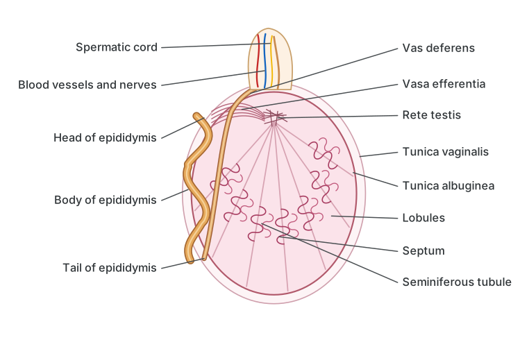
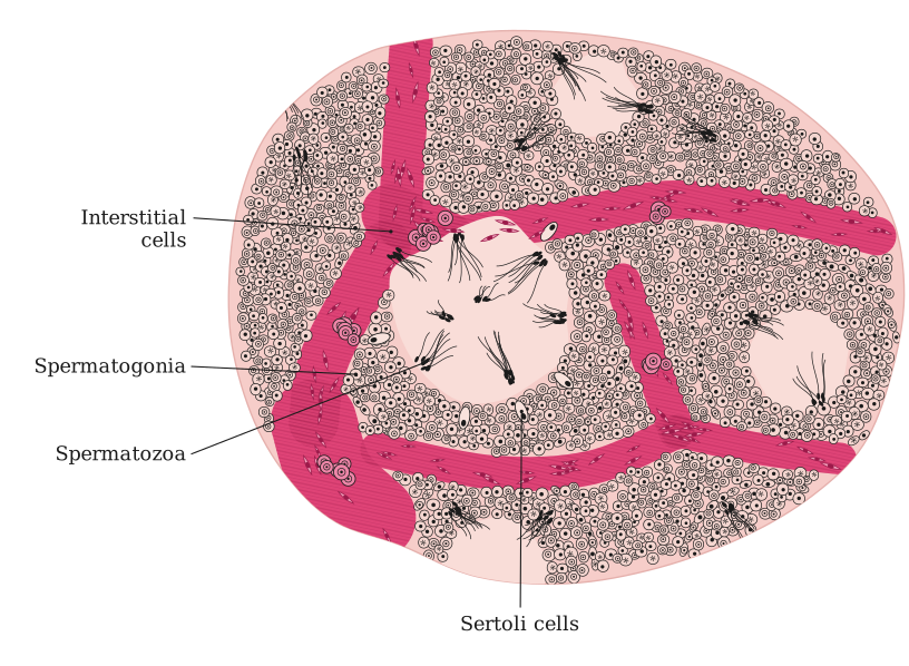
Histology of Testis : Externally the testis is covered by a tough collagenous connective-tissue layer, the tunica albuginea; outside this lies an incomplete peritoneal covering, the tunica vaginalis, and internally, a thin, vascular and membranous layer, the tunica vasculosa. Fibrous septa arising from the tunica albuginea divide each testis internally into about 200–300 compartments called testicular lobules, each containing 1–4 highly coiled seminiferous tubules. Every seminiferous tubule is lined internally by cuboidal germinal epithelial cells (spermatogonia) along with a smaller number of large, pyramidal Sertoli (sustentacular) cells. The germinal epithelial cells undergo spermatogenesis to form spermatozoa, while the Sertoli cells provide nourishment to the developing sperm; the successive stages of spermatogenesis — spermatogonia (2n), primary spermatocyte (2n), secondary spermatocyte (n), spermatid (n) and sperm (n) — can all be seen within the tubule wall. Lying in the loose connective tissue between the seminiferous tubules are the interstitial cells or Leydig cells, which secrete the male hormone, androgen or testosterone.
Drawn by us to help you understand the concept clearly, and verified to make sure it's accurate. For exams, practice from your textbook's own diagram.
What this figure shows. The testis opened lengthwise: the tough tunica albuginea, the septa dividing it into lobules packed with coiled seminiferous tubules, the network of the rete testis at the back, the vasa efferentia leading into the head, body and tail of the epididymis, and the vas deferens leaving it; the sectional view of the pelvis beside it places the testis, scrotum, ducts and glands in position. These are the same parts the textbook's Fig. 2.3 labels (tunica vaginal …
Drawn by us to help you understand the concept clearly, and verified to make sure it's accurate. For exams, practice from your textbook's own diagram.
What this figure shows. A transverse section through the testis: several seminiferous tubules cut across, each bounded by a basement membrane and lined by the germinal epithelium whose spermatogonia mature inwards, with the large Sertoli cells among them and bundles of sperm tails pointing into the lumen; in the connective tissue between the tubules lie the interstitial (Leydig) cells that secrete testosterone, all enclose …
Do you know?
- The presence of a peritoneal covering around the testis is an indication of its abdominal origin. …