Biology · Ch 8 — Respiration and Circulation
Heart
Heart
The heart is the circulatory system's central pump — a reddish-brown, hollow, muscular organ roughly the size of a closed fist, weighing about 300 g in males and 250 g in females. It sits in the mediastinum, the space between the two lungs, is broader at its upper end (the base) and tapers to a point at its lower end (the apex), which is tilted slightly to the left and rests just above the diaphragm.
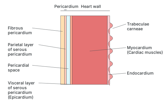
The heart is wrapped in a protective membranous sac, the pericardium, made of an outer fibrous layer and an inner serous layer. The serous pericardium is itself soft, moist and elastic, built of squamous epithelium, and splits into a parietal layer and a visceral layer, separated from each other by a pericardial space filled with about 50 ml of pericardial fluid; this fluid cushions the beating heart against mechanical injury and also keeps it moist and lubricated.
Drawn by us to help you understand the concept clearly, and verified to make sure it's accurate. For exams, practice from your textbook's own diagram.
What this figure shows. A sectioned close-up of the heart wall and its surrounding sac. From outside in, it shows the tough outer fibrous pericardium, then the parietal layer of the serous pericardium, a fluid-filled pericardial space between the two serous layers, the visceral layer of serous pericardium (which is also the heart's own outer covering, the epicardium), the thick middle layer of cardiac muscle (myocardium, shown with its internal ridges, the trabeculae carneae) and the thin innermost lining, the endocardium. It illustrates how the pericardial fluid in the spac …
Heart wall : The heart wall itself, being mesodermal in origin, has three layers: an outer epicardium (a single thin layer of flat squamous epithelium on a basement membrane, which is also the visceral layer of the serous pericardium), a middle myocardium (a thick layer of cardiac muscle, which does the actual work of contracting and relaxing the heart), and an inner endocardium (another single thin squamous layer). The epicardium and endocardium are essentially protective, while the myocardium provides the contractile force.
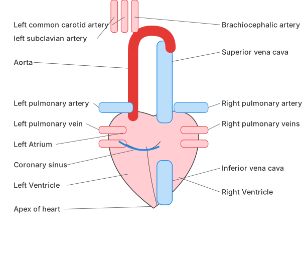
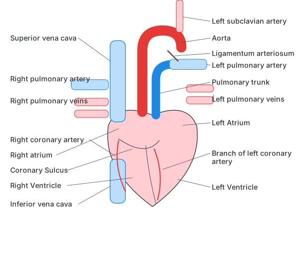
External structure of heart : Externally, the human heart is four-chambered — two upper atria (or auricles) and two lower ventricles — with the atria separated from the ventricles by a groove called the coronary sulcus (or atrioventricular groove), and the two ventricles separated from each other by anterior and posterior inter-ventricular sulci, through which the coronary arteries and veins run. On the front of the heart, the pulmonary trunk arises from the right ventricle (splitting into right and left pulmonary arteries) and the aorta arises from the left ventricle; the aorta itself divides into an ascending part, an aortic arch and a descending part, and the aortic arch gives off three branches — the brachiocephalic (innominate) artery, the left common carotid artery, and the left subclavian artery. A fibrous cord, the ligamentum arteriosum, connects the pulmonary trunk to the aortic arch — a leftover remnant of an embryonic vessel, the ductus arteriosus. On the back of the heart, the superior and inferior vena cava open into the right atrium, while four pulmonary veins open into the left atrium.
Drawn by us to help you understand the concept clearly, and verified to make sure it's accurate. For exams, practice from your textbook's own diagram.
What this figure shows. A back-view diagram of the whole heart. It shows the left atrium receiving the left pulmonary artery and left pulmonary vein, the aorta arching over the top with the left common carotid artery and left subclavian artery branching from it, the coronary sinus running along a groove near the left ventricle, and the pointed apex of the heart at the bottom. On the right side it shows the superior and inferior vena cava opening in near the right atrium, the right pulmonary artery and veins, the right ventricle, and the brachio …
Drawn by us to help you understand the concept clearly, and verified to make sure it's accurate. For exams, practice from your textbook's own diagram.
What this figure shows. A front-view diagram of the whole heart. On the right, it shows the superior and inferior vena cava, the right pulmonary artery and veins, the right coronary artery, the right atrium and, below the coronary sulcus groove, the right ventricle. On the left, it shows the left subclavian artery, the aorta, the ligamentum arteriosum (a fibrous cord linking the pulmonary trunk to the aortic arch, left over from an embryonic blood vessel), the pulmonary trunk splitting toward the lungs, the left pulmonary artery and veins entering the left atrium, a branch of the left co …
Drawn by us to help you understand the concept clearly, and verified to make sure it's accurate. For exams, practice from your textbook's own diagram.
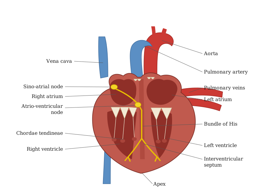
What this figure shows. A front-section cutaway through the heart exposing all four chambers at once. On the right side, the superior vena cava (guarded nearby by the pulmonary semilunar valve on the outflow side) opens into the right atrium, which connects to the right ventricle through the tricuspid valve; the ventricle's inner wall is ridged with columnae carnae, and its valve flaps are tied down by chordae tendinae anchored to papillary muscles. On the left side, pulmonary veins open into the left atrium, which connects to the left ventricle through the bicuspid (mitral) valve, also anchored by chordae tendinae and papillary muscles. The interventricular septum is shown separating the two thick-walled ventricles, with the pulmo …
Internal structure of heart :
Atria : These are thin-walled receiving chambers, separated from each other by the inter-auricular septum, which carries an oval depression, the fossa ovalis — a remnant of an embryonic opening called the foramen ovalis. The superior vena cava, inferior vena cava and coronary sinus all open into the right atrium; the opening of the inferior vena cava is guarded by a Eustachian valve, and the coronary sinus opening by a Thebesian valve. The four pulmonary veins open into the left atrium without any valves at all. Each atrium connects to the ventricle on its own side through an atrioventricular opening guarded by a cuspid valve — the tricuspid valve on the right, and the bicuspid (or mitral) valve on the left — and all of the heart's valves work to keep blood flowing in one direction only, stopping any backflow.
Ventricles : These are the thicker-walled, more muscular pumping chambers below, separated from each other by the interventricular septum; the left ventricle's wall is roughly three times thicker than the right ventricle's, because it has to pump blood at higher pressure all the way around the body, while the right only pumps to the nearby lungs. The inner surface of each ventricle is ridged with muscular projections called columnae carnae (trabeculae carnae), and inelastic fibres called chordae tendinae run from the tricuspid and bicuspid valves down to small projections of ventricular muscle called papillary muscles, anchoring the valve flaps and controlling how they open and close so they don't flip backward under pressure. The right ventricle opens into the pulmonary trunk and the left ventricle into the aorta, each guarded by three semilunar valves that stop blood flowing backward into the ventricles once it has been pumped out.
Pumping action of heart : The heart acts as the main pumping organ of the circulatory system. The pumping action is brought about by a rhythmic contraction and relaxation of the cardiac muscles. Contraction of the heart muscles is systole and relaxation is diastole. A single systole followed by a diastole makes one heart beat; the heart beats 70 to 72 times per minute (the heart rate). During each beat the ventricles pump about 70 ml of blood (the stroke volume), so the heart pumps about 72 x 70 ml = 5040 ml, roughly 5 litres, of blood per minute - the cardiac output.
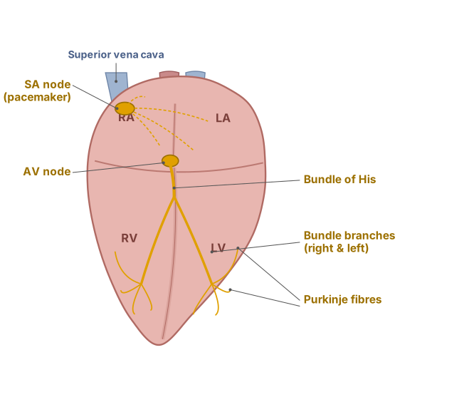
Conducting tissue of heart : The heart's ability to beat on its own, without any outside nerve signal, is called being myogenic, and its ability to set its own rhythm is called auto-rhythmicity; both properties come from a specialised patch of cardiac muscle called the nodal (conducting) tissue.
Drawn by us to help you understand the concept clearly, and verified to make sure it's accurate. For exams, practice from your textbook's own diagram.
What this figure shows. A diagram of the heart's built-in electrical wiring. It starts at the sino-atrial (SA) node in the upper wall of the right atrium (the pacemaker), from where the impulse spreads across both atria and also reaches the atrio-ventricular (AV) node near the base of the interatrial septum in the right atrial wall. From the AV node, the atrioventricular bundle (bundle of His) carries the impulse through the interventricular septum and splits into right and left bundle branches, one running down into each ventricle, ending in a fine branching network called Purkinje fibres that spreads the impulse …
…