Biology · Ch 8 — Respiration and Circulation
Thrombocytes / Platelets
Thrombocytes / Platelets
Thrombocytes, or platelets, are not whole cells but small cell fragments, broken off from large bone-marrow cells called megakaryocytes; they are oval, lack a nucleus, and are much smaller than either RBCs or WBCs. Normal platelet count is about 2.5-4.5 lakh per mm3 of blood. If the count drops below normal, the condition is called thrombocytopenia, which causes internal bleeding (haemorrhage) because clotting cannot proceed properly. Platelets carry out two key jobs in stopping blood loss: they secrete platelet factors essential for clotting, and they physically seal small ruptures in blood vessels by clumping together into a platelet plug (thrombus); they also secrete serotonin, which acts locally as a vasoconstrictor to narrow the damaged vessel and reduce blood flow to it.
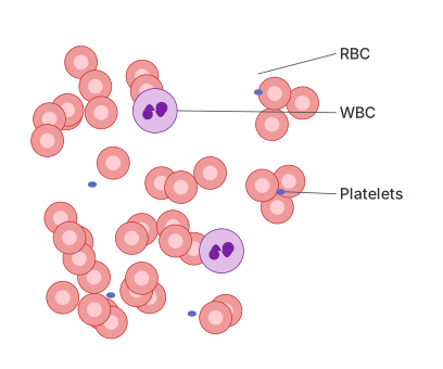
Drawn by us to help you understand the concept clearly, and verified to make sure it's accurate. For exams, practice from your textbook's own diagram.
What this figure shows. A microscope-field view of a stained blood smear laid out to show the relative appearance and abundance of the three formed elements together: numerous small, biconcave, nucleus-free red blood cells filling most of the field; a much smaller number of larger, nucleated white blood cells scattered among them; and tiny, irregularly shaped, anucleate platelet fragments dotted around, illustrating just how much more numerous RBCs …
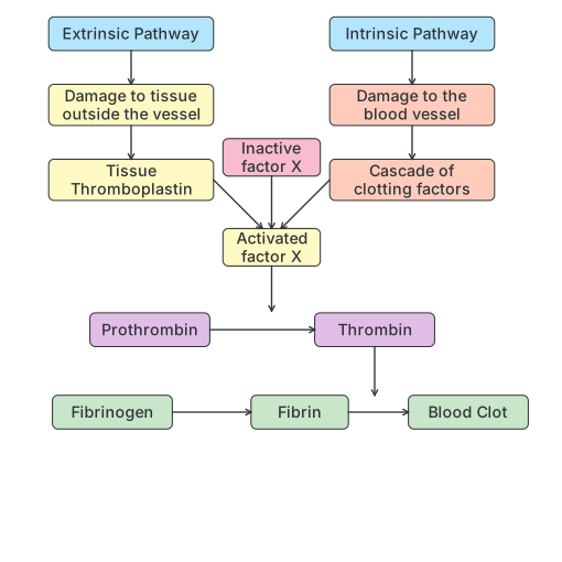
Blood Clotting/ Coagulation of blood : Blood clotting (coagulation) is the process that converts liquid blood into a solid mass, and it can be triggered in two ways: by blood coming into contact with any foreign surface (the intrinsic pathway) or by contact with damaged tissue (the extrinsic pathway). Both pathways work through a step-by-step cascade of interacting clotting factors — there are twelve in total, numbered I to XIII (factor VI is not used). In the extrinsic pathway, damage to tissue outside a vessel releases tissue thromboplastin; in the intrinsic pathway, damage to the vessel wall itself sets off the cascade directly. Both pathways converge on activating clotting factor X; thromboplastin also helps form the enzyme prothrombinase, which inactivates heparin (the natural anticoagulant) and converts inactive prothrombin into active thrombin. Thrombin then converts the soluble plasma protein fibrinogen into insoluble fibrin, which forms a tangled mesh that traps platelets and other blood cells, forming the clot.
Observe and Discuss
Observe the flow chart and discuss the process with your friends.
Drawn by us to help you understand the concept clearly, and verified to make sure it's accurate. For exams, practice from your textbook's own diagram.
What this figure shows. A flowchart showing how liquid blood is converted to a solid clot through two pathways that converge on the same final steps. The extrinsic pathway begins when tissue outside a blood vessel is damaged, releasing tissue thromboplastin; the intrinsic pathway begins when the vessel wall itself is damaged, setting off a cascade of clotting factors circulating in the blood. Both pathways lead to the activation of clotting factor X, which converts prothrombin into the active enzyme thrombin. Thrombin then converts the soluble plasma protein fibrinogen into insoluble fibrin threads, which weave into a mesh that …
Curiosity
- What is blood clotting? How and when does it occur? …