Biology · Ch 8 — Respiration and Circulation
Mechanism of respiration
Mechanism of respiration
Respiration is a biological process involving the exchange of gases between the atmosphere and the lungs, and it results in the formation of ATP. It includes the following processes:
A. Breathing
B. External respiration
C. Internal respiration
D. Cellular respiration
Try This
Count the number of breaths you take in the following situations:
- after a good night's sleep;
- during a vigorous activity (running, climbing stairs, etc.);
- after the vigorous activity. Do you find any difference in the count?
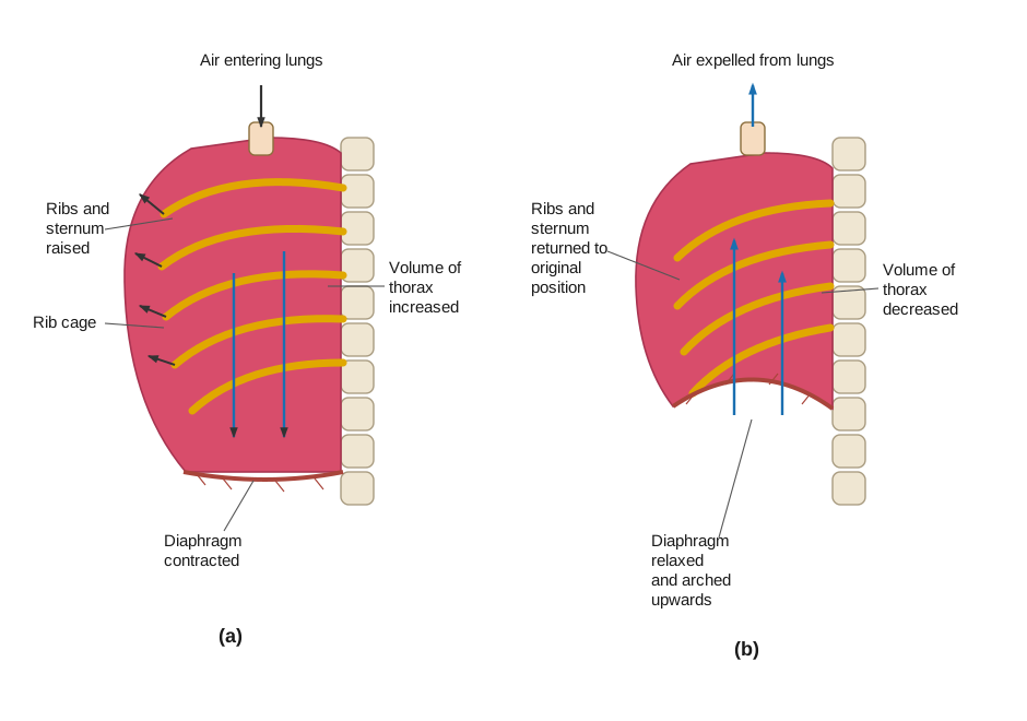
A. Breathing : This is the purely physical, mechanical step of moving air in and out of the lungs, driven by the ribs, sternum, intercostal muscles and diaphragm working together. During inspiration, the diaphragm contracts and flattens downward while the external intercostal muscles contract, pulling the ribs and sternum up and outward; this enlarges the thoracic cavity, drops the pressure inside the lungs below atmospheric pressure, and air rushes in to equalise it — inspiration is therefore an active, muscle-driven process. During expiration, the diaphragm relaxes back into its dome shape and the intercostal muscles relax, letting the rib cage move down and inward; this shrinks the thoracic cavity, raises the pressure inside the lungs above atmospheric pressure, and air is pushed back out. One inspiration plus one expiration makes one breath
Drawn by us to help you understand the concept clearly, and verified to make sure it's accurate. For exams, practice from your textbook's own diagram.
What this figure shows. A two-panel diagram contrasting the two phases of one breath. In panel (a), inspiration, the diaphragm is shown contracting and flattening downward while the rib cage is pulled up and outward by the external intercostal muscles, enlarging the thorax so that falling internal pressure draws air in. In panel (b), expiration, the diaphragm is shown relaxing back into its dome shape and the rib cage moving down and inward as the intercostal muscles relax, shrinking the thorax so that ri …
Pulmonary volumes and capacities (Normal values)
Lung Volumes :
Tidal volume (T.V.) : the volume of air inspired or expired during normal breathing - 500 ml.
Inspiratory reserve volume (IRV) : the maximum (extra) volume of air inspired during forced breathing in addition to T.V. - 2000 to 3000 ml.
Expiratory reserve volume (ERV) : the maximum volume of air expired during forced breathing after a normal expiration - 1000 to 1100 ml.
Dead space (DS) : the volume of air present in the respiratory tract (from the nose to the terminal bronchioles) but not involved in gaseous exchange - 150 ml.
Residual volume (RV) : the volume of air that remains in the lungs and the dead space even after maximum expiration - 1100 to 1200 ml.
Lung capacities :
Total Lung capacity : the maximum amount of air the lungs can hold after a maximum forceful inspiration - 5200 to 5800 ml.
Vital capacity (VC) : the maximum amount of air that can be breathed out after a maximum inspiration; the sum total of TV, IRV and ERV - 4100 to 4600 ml.
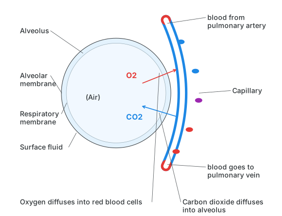
B. External respiration/ Exchange of gases at the alveolar level : This is the actual gas exchange between alveolar air and the blood in the pulmonary capillaries, made possible because the alveolar wall and the capillary wall are both single layers of thin squamous epithelium that together form the respiratory membrane. Gases simply diffuse from wherever their partial pressure is higher to wherever it is lower, until the two sides equalise. Blood arriving in the pulmonary capillaries carries carbon dioxide at about 45 mmHg while the alveolar air sits at about 40 mmHg, so CO2 diffuses from blood into the alveolus; the same blood carries oxygen at about 40 mmHg while alveolar air is at about 104 mmHg, so O2 diffuses from the alveolus into the blood.
Drawn by us to help you understand the concept clearly, and verified to make sure it's accurate. For exams, practice from your textbook's own diagram.
What this figure shows. A close-up of one alveolus pressed against a surrounding blood capillary, showing blood arriving from the pulmonary artery. Oxygen (higher partial pressure in the alveolar air) is shown diffusing across the shared respiratory membrane — the alveolar lining, the fused basement membrane and the capillary wall, plus a thin surface fluid film — into the red blood cells, while carbon dioxide (higher partial pressure in the blood) diffuses the opposite way into the alveolar air, after which the now oxygen-riche …
Use your brain power
Why is gas exchange in the alveolar region very rapid?
C. Internal respiration : The two main components of blood involved in the transport of the respiratory gases CO2 and O2 are the RBCs and the plasma.
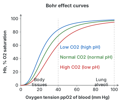
i. Transport of oxygen : For oxygen, only about 3% travels dissolved directly in the plasma; the remaining 97% is carried bound to haemoglobin (Hb) inside red blood cells, which acts as the respiratory pigment. Each haemoglobin molecule has four iron (Fe++) sites, and each can bind one oxygen molecule, so one Hb molecule can carry up to four O2 as oxyhaemoglobin (Hb + 4O2 → Hb(4O2)); at the tissues this dissociates again to release the oxygen (Hb(4O2) → Hb + 4O2). How saturated haemoglobin gets depends on the surrounding oxygen tension (ppO2): saturation reaches 95-97% at the high ppO2 (about 100 mmHg) found in the alveoli, and drops off as ppO2 falls — at only 30 mmHg, saturation falls to about 50%. This relationship, plotted as percentage saturation against ppO2, gives the S-shaped oxyhaemoglobin dissociation curve, which shifts further to the right (meaning haemoglobin releases oxygen more readily at any given ppO2) when H+ concentration rises, CO2 tension rises, temperature rises, or 2,3-DPG (a by-product of glycolysis in red blood cells) rises — exactly the conditions found in active, oxygen-hungry tissue. This rightward shift caused specifically by a change in blood CO2 is called the Bohr effect. A related phenomenon, the Haldane effect, describes how oxyhaemoglobin itself behaves like a weak acid, lowering blood pH as its H+ ions increase and driving bicarbonate ions to break back down into water and CO2. In practical terms, this means that in the alveoli, where ppO2 is high and ppCO2 is low, haemoglobin binds oxygen readily, while in the tissues, where ppO2 is low and ppCO2 is high, haemoglobin holds onto much less oxygen and releases it for diffusion into the cells.
Drawn by us to help you understand the concept clearly, and verified to make sure it's accurate. For exams, practice from your textbook's own diagram.
What this figure shows. A graph plotting the percentage saturation of haemoglobin with oxygen (y-axis) against the oxygen tension, ppO2, of the blood in mmHg (x-axis). The line has a shallow start, a steep middle rise and a flattened top, giving it an S (sigmoid) shape rather than a straight line — showing that haemoglobin is nearly fully saturated (95-97%) at the high ppO2 found in the lungs but gives up oxygen readily once ppO2 drops to the lower levels found in active tissue. The curve is drawn shifting further to the right when hydrogen-ion concentration, carbon-dioxide tension, temperature or 2,3-DPG rise, reflect …
Carbon monoxide poisoning
The affinity of haemoglobin for carbon monoxide is about 250 times more than for oxygen: haemoglobin's affinity for carbon monoxide is roughly 250 times greater than for oxygen, so even small amounts of CO form a stable compound, carboxyhaemoglobin, that blocks haemoglobin from carrying oxygen at all — starving the tissues of oxygen even though the blood is technically 'full'.
Use your brain power
While working with the car engine in a closed garage, John suddenly felt dizzy and fainted. What is the possible reason?
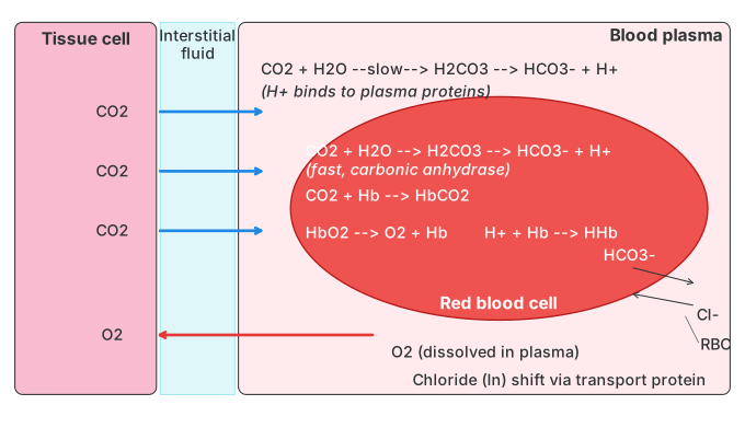
ii. Transport of CO2 : Carbon dioxide is readily soluble in water and is transported by the RBCs and the plasma in three different forms.
a. By plasma in solution form (7 %) : About 7 % simply dissolves in the plasma as carbonic acid (CO2 + H2O ⇌ H2CO3). …
Drawn by us to help you understand the concept clearly, and verified to make sure it's accurate. For exams, practice from your textbook's own diagram.
What this figure shows. A diagram of a tissue cell bathed in interstitial fluid next to a red blood cell inside a capillary. It traces oxyhaemoglobin (HbO2) releasing its oxygen, which diffuses out through the plasma and interstitial fluid into the tissue cell, while carbon dioxide produced by the tissue diffuses inward — some staying dissolved in plasma, some entering the red blood cell to be converted by carbonic anhydrase into carbonic acid and then bicarbonate plus hydrogen ions (with chloride ions shifting into the cell to balance the charge lost as bicarbonate leaves), and some binding haemoglobin's globin …
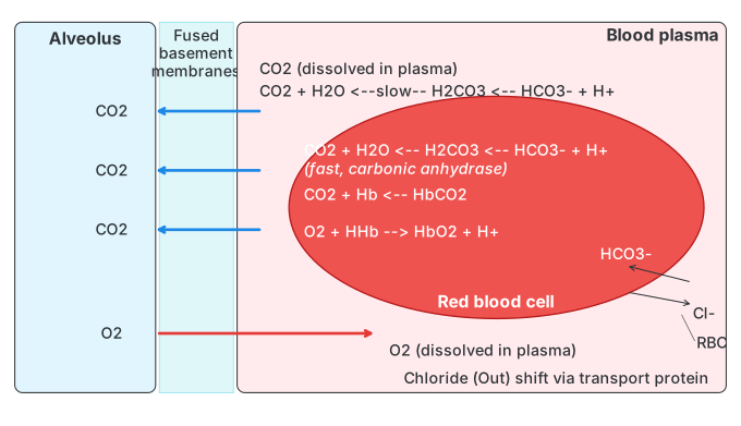
Drawn by us to help you understand the concept clearly, and verified to make sure it's accurate. For exams, practice from your textbook's own diagram.
What this figure shows. The mirror-image diagram to Fig. 8.8, set at the alveolus instead of the tissue. It shows bicarbonate ions and hydrogen ions recombining inside the red blood cell (helped by carbonic anhydrase) to regenerate carbon dioxide and water, which then diffuses out into the alveolar air, while chloride ions shift back out of the cell (the reverse of the tissue-level chloride shift) and oxygen from the alveolar air diffuses in to bind reduced haemoglobin, reforming oxyhaemoglobin ready t …