Biology · Ch 8 — Respiration and Circulation
Regulation of Breathing
Regulation of Breathing
Breathing is not something we have to think about — it runs automatically under dual control, nervous and chemical, and is only loosely under voluntary override. An adult breathes about 12 times a minute at rest, while a newborn breathes far faster, about 44 times a minute. The steady, involuntary rhythm is set by groups of neurons in the pons and medulla of the brainstem, together called the respiratory centres, which control both the rate and depth of breathing. These are organised into a dorsal group of neurons in the medulla (the inspiratory centre), a ventro-lateral group in the medulla (handling both inspiration and expiration), and a pneumotaxic centre in the pons, which mainly limits the length of inspiration and is also active during slow-wave and REM sleep; an apneustic centre in the medulla works against the pneumotaxic centre and is involved in controlling non-REM sleep and wakefulness.
Drawn by us to help you understand the concept clearly, and verified to make sure it's accurate. For exams, practice from your textbook's own diagram.
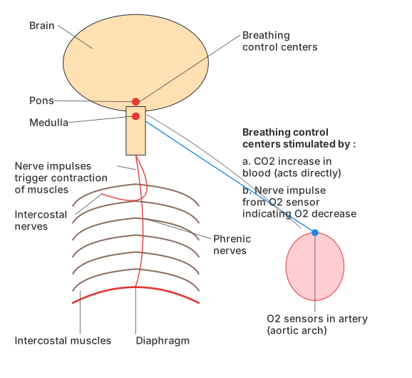
What this figure shows. A flow diagram centred on the brain's breathing control centres. Two inputs feed into these centres: a rise in blood carbon dioxide, which acts on them directly, and a nerve impulse from oxygen sensors in the aortic arch signalling a fall in oxygen. From the control centres, outgoing nerve impulses travel down the phrenic nerves to the diaphragm and the intercostal nerves to the intercostal muscles, triggering the muscle contractions that produce a breath. The diagram places these co …
The respiratory centres are driven mainly by chemical signals: a rise in blood CO2 acts on them directly, while a fall in blood O2 is detected by O2 sensors in the aortic arch, which send a nerve impulse to the centres instead. Once triggered, the centres send nerve impulses down the phrenic nerves to the diaphragm and the intercostal nerves to the intercostal muscles, causing them to contract and produce a breath.
Breathing depth and rhythm are additionally fine-tuned by a reflex loop: as the lungs expand during inspiration to a critical point, stretch receptors in the lung tissue are triggered and send impulses along the vagus nerve to the expiratory centre, which in turn sends inhibitory signals to the inspiratory centre. This relaxes the inspiratory muscles and lets expiration follow; as air leaves and the lungs deflate, the stretch receptors fall silent, releasing the inspiratory centre from inhibition so a new breath can begin. This loop is the Hering-Breuer reflex, and besides pacing normal breathing it also protects the lungs from over-inflating to the point of bursting. …