Biology · Ch 8 — Respiration and Circulation
Lymphatic System
Lymphatic System
The lymphatic system is made up of lymph (the fluid itself), lymphatic vessels, and a set of associated organs and tissues. Lymph, literally 'clear water', is a fluid connective tissue whose composition is very close to blood plasma, except that it lacks red blood cells, platelets and some of the larger plasma proteins. It forms when fluid from the intercellular (interstitial) spaces of body tissue seeps into blind-ended lymphatic capillaries; from there it travels through progressively larger lymphatic vessels — picking up filtering along the way at lymph nodes — until it is finally emptied back into the blood via the thoracic duct and the right lymphatic duct, both of which drain into the subclavian vein.
Internet my friend
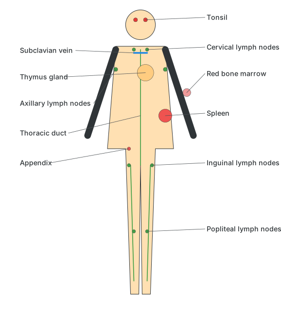
Location of lymph nodes in the human body.
Drawn by us to help you understand the concept clearly, and verified to make sure it's accurate. For exams, practice from your textbook's own diagram.
What this figure shows. A whole-body diagram marking the location of the major lymphatic organs and lymph node clusters: the thymus gland and cervical lymph nodes near the neck/chest, the thoracic duct running up through the torso to drain into the subclavian vein, axillary lymph nodes under the arm, the spleen and red bone marrow, the appendix and a tonsil as examples of lymphoid tissue, and inguinal and popliteal lymph node clusters in the groin and behind the knee — showing that lymphatic tissue and drainage points are sprea …
Lymph nodes and lymphoid tissue are distributed widely through the body rather than concentrated in one place: examples include the thymus gland, tonsils and cervical lymph nodes near the head and neck, axillary lymph nodes under the arms, the appendix, the spleen and red bone marrow, and inguinal and popliteal lymph nodes in the groin and behind the knee.
Do you know?
Tonsils in particular are small lymphatic nodules in the pharyngeal region — normally five of them, positioned to intercept inhaled and ingested foreign material — and their inflammation, tonsillitis, caused by viral or bacterial infection, produces a sore throat, fever, swollen lymph nodes, nasal congestion, difficulty swallowing and headache; viral tonsillitis usually resolves on its own, while bacterial tonsillitis needs antibiotics, and cases that do not respond to treatment may need surgical removal (tonsillectomy).
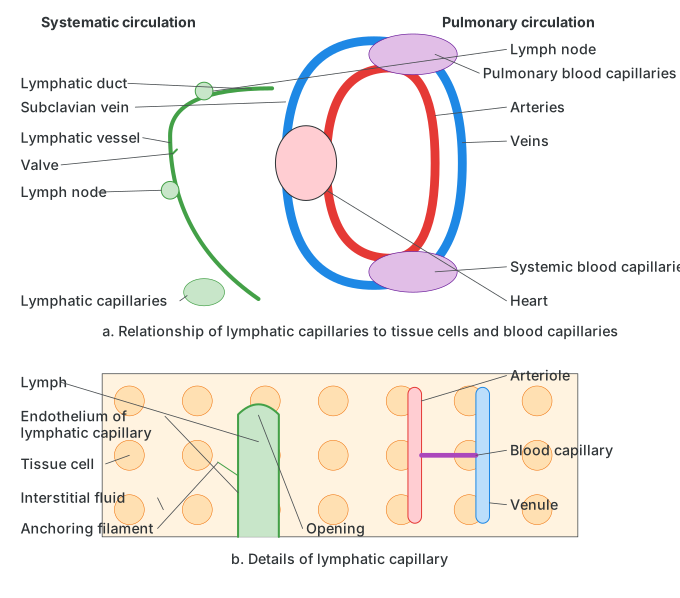
A lymphatic capillary works because of how its wall is built: a single layer of overlapping endothelial cells held in place by anchoring filaments, creating flap-like openings that let interstitial fluid (and larger particles and proteins that have leaked from blood capillaries) enter easily but close up to stop that fluid leaking back out once inside. This lets lymphatic capillaries running alongside the systemic and pulmonary blood capillaries continually drain away excess interstitial fluid, route it through the chain of lymph nodes (where it is filtered and checked for pathogens) and larger valved lymphatic vessels, and finally return it to general circulation via the subclavian vein — completing the fluid-balance role that keeps the volume of interstitial fluid around the body's cells stable, and closing the loop this chapter opened in section 8.7 on how a complex body moves material between its parts.
Drawn by us to help you understand the concept clearly, and verified to make sure it's accurate. For exams, practice from your textbook's own diagram.
What this figure shows. A two-part diagram relating the lymphatic network to the blood circulation. Part (a) shows lymphatic capillaries running alongside the systemic and pulmonary blood capillaries near the heart, collecting fluid and passing it through a chain of lymph nodes and larger lymphatic vessels (each with internal valves, like veins) up to a lymphatic duct that empties into the subclavian vein, rejoining the bloodstream. Part (b) zooms into a single lymphatic capillary sitting between a tissue cell and a blood capillary, showing its wall of overlapping endothelial cells held by anchoring filaments that create one-way openings — letting interstitial fluid (and the blood plasma proteins/fluid that leaked fr …
Activity
- Prepare a chart of the different types of heart muscles and their functions (atrial muscle, ventricular muscle and the specialised nodal or conducting muscle fibres - SA node, AV node, bundle of His, Purkinje fibres).
- Differentiate between the functioning of the heart in the frog and in the human.
| Human Heart | Frog Heart |
|---|---|
| Four chambers: two atria and two ventricles | Three chambers: two atria and one ventricle |