Biology · Ch 8 — Respiration and Circulation
White blood corpuscles / Leucocytes
White blood corpuscles / Leucocytes
Leucocytes, or white blood corpuscles, are the blood's mobile defence force. Unlike RBCs, they are colourless, nucleated, larger, and capable of amoeboid movement, which lets them squeeze out through capillary walls into surrounding tissue by a process called diapedesis whenever they are needed to fight infection. A healthy adult has, on average, 5000-11000 WBCs per mm3 of blood. A drop below about 4000 is called leucopenia (seen in HIV/AIDS, tuberculosis, or after radiation exposure or shock); a temporary rise is leucocytosis, typically caused by infection, and also seen normally during pregnancy and in newborns; an uncontrolled, sustained rise is leukemia, a type of blood cancer. WBCs, in short, exist mainly for protection rather than transport.
Types of WBCs : WBCs are irregular, nucleated and 'polymorphic' (they occur in variable forms), and fall into two broad groups: granulocytes and agranulocytes.
A. Granulocytes : Granulocytes have a visibly granular cytoplasm and a lobed nucleus, which is why they are also called polymorphonuclear leucocytes (PMN cells). They develop from myeloid stem cells and, once formed, no longer divide; together they make up about 72% of all WBCs. Their granules are actually secretory vesicles holding various enzymes and secretions, and depending on how these granules take up different stains, granulocytes split into three types.
Think about it
What are the granules in granulocytes?
a. Neutrophils : Neutrophils, about 70 % of all WBCs, have very fine, evenly distributed granules that stain with neutral dyes, a nucleus lobed into 2-7 parts, and the ability to move amoeboidly and carry out phagocytosis, engulfing and destroying pathogens; 'pus' is essentially a mixture of dead neutrophils, damaged tissue and dead microbes.
Think about it
Why and when do the leucocytes perform diapedesis?
b. Basophils / Cyanophils : Basophils, only about 0.5-1% of WBCs, are small, spherical, non-phagocytic cells with a twisted nucleus and a few large granules that stain with basic dyes like methylene blue; they appear mainly during infection and allergic reactions, and secrete heparin, histamine and serotonin.
c. Eosinophils / Acidophils : Eosinophils, about 1-3% of WBCs, have a bilobed nucleus and lysosomal granules that stain red with acidic stains like eosin; their numbers rise in allergic conditions, they destroy antigen-antibody complexes by phagocytosis, show an antihistamine-like effect, and also help detoxify the body by producing antitoxins.
B. Agranulocytes : Agranulocytes, about 28% of WBCs, have cytoplasm without visible granules, develop from lymphoid stem cells, and — unlike granulocytes — can still divide by mitosis; their nuclei are large but not lobed. There are two types. …
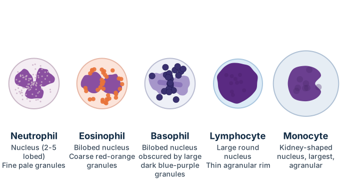
Drawn by us to help you understand the concept clearly, and verified to make sure it's accurate. For exams, practice from your textbook's own diagram.
What this figure shows. A set of five labelled cell drawings comparing the shapes of the different white blood cell types side by side: (a) the neutrophil, with a nucleus divided into several (2-7) lobes and fine, evenly distributed granules; (b) the basophil, smaller and spherical with a twisted nucleus and a few large granules; (c) the eosinophil, with a bilobed nucleus and granules that take up acidic (red) stain; (d) the lymphocyte, the smallest of the five, almost filled by one large spherical nucleus with very little visible cytoplasm; and (e) the monocyte, the largest of the five, with a big bean- or kidney-shaped nucleus. The figure is built to le …