Biology · Ch 8 — Respiration and Circulation
Working mechanism of human heart
Working mechanism of human heart
Cardiac Cycle : The heart's pumping action comes from the rhythmic contraction and relaxation of cardiac muscle. Contraction is called systole, and relaxation is called diastole; one systole followed by one diastole makes a single heartbeat, or cardiac cycle, and this whole cycle takes about 0.8 seconds, giving a resting adult heart rate of 70-72 beats per minute.
Within one cardiac cycle, atria and ventricles contract and relax alternately, in three phases.
a. Atrial systole (AS) : In atrial systole, once both atria are full of blood (the right atrium receiving deoxygenated blood, the left receiving oxygenated blood), the resulting pressure on the atrial wall excites the SA node into generating a cardiac impulse; the atrial muscle then contracts, pumping blood into the ventricles, while the Eustachian and Thebesian valves stop blood being pushed backward into the veins and coronary sinus. Atrial systole lasts about 0.1 second, followed by atrial diastole lasting about 0.7 second.
b. Ventricular systole (VS) : In ventricular systole, the impulse that started at the SA node reaches and excites the AV node, which passes it on through the bundle of His to the Purkinje fibres, spreading it across the ventricular walls and causing them to contract; the right ventricle pumps deoxygenated blood into the pulmonary trunk while the left pumps oxygenated blood into the aorta, and during this phase the tricuspid and bicuspid (cuspid) valves close to stop blood flowing back up into the atria — producing the first heart sound, 'lubb'. Ventricular systole lasts about 0.3 second, followed by ventricular diastole lasting about 0.5 second, during which the semilunar valves close to stop blood flowing back from the pulmonary trunk and aorta into the ventricles — producing the second heart sound, 'dub'. For about 0.4 second within this sequence, both the atria and ventricles are relaxed at the same time, a state called joint diastole (or complete diastole). Adding these overlapping phases together accounts for the full 0.8-second cardiac cycle.
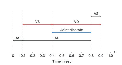
Drawn by us to help you understand the concept clearly, and verified to make sure it's accurate. For exams, practice from your textbook's own diagram.
What this figure shows. A timeline graph running from 0 to 0.8 seconds along the x-axis, tracking the state of the atria and ventricles through one full heartbeat. It marks atrial systole (AS) lasting about 0.1 second followed by atrial diastole (AD), and separately marks ventricular systole (VS) lasting about 0.3 second followed by ventricular diastole (VD); the final stretch of the timeline, where both atria and ventricles are relaxed together, is labelled joint diastole. Reading the bands stacked against the timeline shows how the 0.8-second cardiac cycle is built up fro …
Total volume of blood pumped during one ventricular systole is called the stroke volume (SV); it is approximately 70 ml.
Cardiac output (CO) : It is the volume of blood pumped out per minute. For a normal adult human being it is calculated as CO = SV x HR = 70 x 72 = 5040 ml/min.
Regulation of cardiac activity …