Biology · Ch 8 — Respiration and Circulation
Human Respiratory System
Human Respiratory System
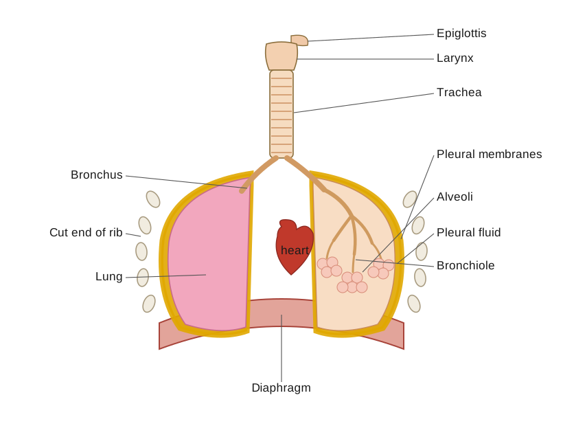
In humans, the respiratory system has two parts: an upper part (external nares, nasal cavities, internal nares, nasopharynx, nose, throat and associated structures) and a lower part (larynx, trachea, bronchi, bronchioles and lungs), and together they bring in air, pass it to the lungs, and hand off the actual exchange of gases to the blood.
Drawn by us to help you understand the concept clearly, and verified to make sure it's accurate. For exams, practice from your textbook's own diagram.
What this figure shows. A whole-airway diagram tracing the path air takes from the nostril through the nasal cavity, into the pharynx and mouth region, down through the larynx and trachea, which then splits at the right main bronchus into a branching tree inside the right lung (with its three lobes) and the smaller left lung (two lobes). The diaphragm is shown as a dome-shaped sheet of muscle beneath the lungs and the ribs enclosing the thoracic cage around them. An inset magnifies a single alveolus — a thin-walled air sac wrapped in a mesh of blood capi …
Nose : Air enters through the external nares (nostrils), a pair of slit-like openings, into the nasal cavity, which is split into a right and left chamber by a cartilage partition. Each chamber is further divided front-to-back into three regions: the vestibule near the nostrils, whose skin bears hairs that filter out dust and larger particles from incoming air; the respiratory part (or conditioner), a thin, highly vascular middle region that warms and moistens the air; and the olfactory or sensory chamber at the top, lined with olfactory epithelium that detects smell.
Pharynx : From the nasal chambers, air passes into the pharynx, which has three regions: the nasopharynx (uppermost, continuous with the nasal chamber), the oropharynx (a shared passage for both food and air, separated from the nasopharynx by the palate bone), and the laryngopharynx below that. The pharynx also carries a set of lymphoid organs, the tonsils, which help guard against pathogens entering via the mouth or nose.
Find out
- Kavya underwent a surgical procedure called rhinoplasty. What could have been the reason for such a surgery? On which part of the body is it carried out?
- What is the role of tonsils in our body? How many pairs of tonsils do we have?
Do you know?
- Shreyas choked while eating dinner. How can you help him? What is the immediate help that can be given to him?
- What is the role of the epiglottis?
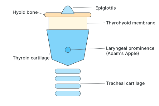
Larynx : Beyond the pharynx is the larynx, or voice box — a hollow tube whose wall is built of cartilage plates held together by membranes and muscles, and which contains the true vocal cords, a pair of elastic folds. Sound is produced as air passes between the vocal cords, with the tongue, teeth, lips and nasal cavity modulating it into speech. The larynx opens into the laryngopharynx through a narrow slit called the glottis, and this opening is guarded by a leaf-shaped flap, the epiglottis, which folds down during swallowing to stop food entering the trachea.
Drawn by us to help you understand the concept clearly, and verified to make sure it's accurate. For exams, practice from your textbook's own diagram.
What this figure shows. A side-on diagram of the voice box built up from cartilage plates joined by membranes and muscle. From top to bottom it shows the hyoid bone, the thyrohyoid membrane connecting it to the thyroid cartilage (whose forward projection is the laryngeal prominence, popularly the Adam's apple), the leaf-shaped epiglottis flap sitting above the opening into the larynx, and the tracheal cartilage rings continuing down into the windpipe. It illustrates how the epiglottis …
Trachea (wind pipe) and Bronchi : The trachea (windpipe) is a 10-12 cm tube running down the neck in front of the oesophagus into the thoracic cavity. Its wall is held open by 16-20 incomplete, C-shaped rings of cartilage that stop it collapsing, and it is lined internally with ciliated, mucus-producing pseudostratified epithelium that traps unwanted particles before they reach the lungs. In the middle of the thoracic cavity, the trachea divides into a right and a left primary bronchus, each also supported by C-shaped cartilage rings; these divide further into secondary and tertiary bronchi, then into terminal bronchioles that finally end in clusters of alveoli. …