Q.Describe the histology of 'human testis'. Write a note on human sperm. OR With the help of a neat and labelled diagram describe the anatomy of 'human eye'. Explain the mechanism of vision.
You're viewing a preview — the full solution, concept, methods & PYQ mapping are locked.
Start your 14-day free trial to unlock the full solution →Testis histology: seminiferous tubules with spermatogenic cell layers + supportive Sertoli cells, interstitial Leydig cells (secrete testosterone) between tubules; sperm has an acrosome-capped head, mitochondria-packe...
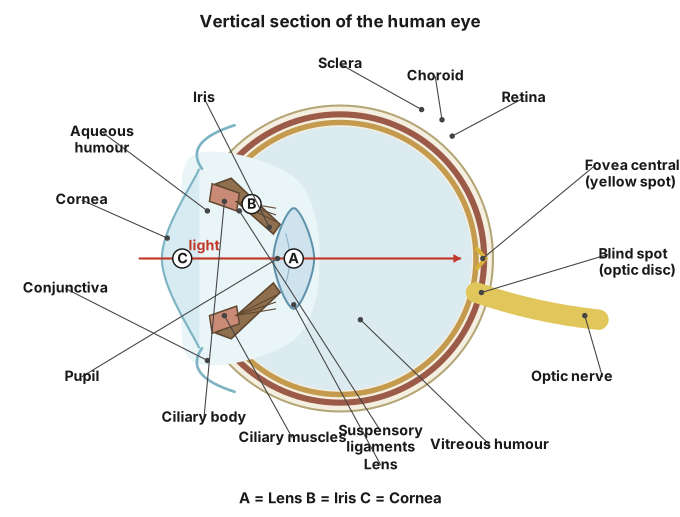
Testis = tubules (sperm-forming + Sertoli cells) + interstitial Leydig cells; sperm = head+middle piece+tail. OR Eye = 3-layered wall + lens, focusing light onto the retina for phototransduction and cortical interpretation.
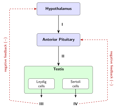
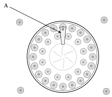
Histology of human testis: Each testis is packed with numerous highly coiled seminiferous tubules, bundled into lobules by connective-tissue septa. The wall of each tubule contains two cell types: spermatogenic cells, arranged in progressively maturing layers from the tubule periphery (spermatogonia) inward through primary and secondary spermatocytes and spermatids, to mature spermatozoa released at the lumen; and tall Sertoli (nurse) cells interspersed among them, which physically support, nourish and protect the developing sperm cells and secrete regulatory substances such as androgen-binding protein and inhibin. Between the tubules, in the loose interstitial connective tissue, lie clusters of Leydig (interstitial) cells, which secrete the male sex hormone testosterone. The tubules of each lobule converge into the rete testis, which drains via the vasa efferentia into the epididymis.
Note on human sperm: A mature sperm has three regions — a head (containing a condensed haploid nucleus, capped by the enzyme-filled acrosome that helps penetrate the egg's coverings), a middle piece (packed with spirally-arranged mitochondria that provide ATP for movement), and a tail/flagellum (which propels the sperm by lashing movements). Sperm are non-motile in the testis and only gain full motility and fertilising ability later, during passage through the epididymis and after capacitation in the female reproductive tract. …
Unlock everything free for 14 days
- Full step-by-step solutions
- Concept-first explanations
- Methods, shortcuts & mistakes
- PYQ mapping + timed mock tests
Full access for 14 days. No credit card required.