Q.Describe, with reference to each stage, the complete life cycle of Entamoeba histolytica.
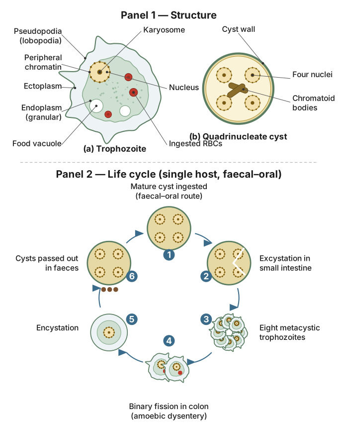
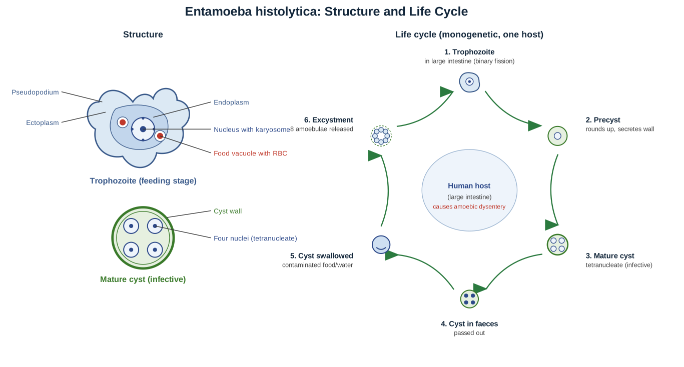
Entamoeba histolytica has one of the simplest life cycles studied in this chapter, requiring only a single host — man — with no intermediate host or vector, though contaminated food, water, unwashed hands and even flies act as passive carriers of its infective stage between hosts.
Stage 1: Ingestion of the mature cyst
Infection begins when a person swallows the mature, resistant quadrinucleate cyst of Entamoeba histolytica, present in food or water contaminated by the faeces of an infected person. The cyst's tough wall protects it as it passes through the acidic environment of the stomach unharmed.
Stage 2: Excystation and the metacystic trophozoite
On reaching the small intestine, digestive juices act on the cyst wall and dissolve it, a process called excystation. This releases a single amoeba which, because the cyst contained four nuclei, almost immediately undergoes cytoplasmic division to give rise to eight small, uninucleate metacystic trophozoites. These newly formed trophozoites then migrate onward from the small intestine to the large intestine (colon), which is their final habitat.
Stage 3: Growth and multiplication of the trophozoite in the colon
In the colon, the metacystic trophozoites grow into full-sized trophozoites — naked, amoeboid cells with a clear outer ectoplasm and granular inner endoplasm, moving and feeding by means of pseudopodia, and each containing a single nucleus with a central karyosome. They feed chiefly on bacteria and cell debris present in the gut lumen, and multiply repeatedly by simple binary fission, building up a colonising population along the mucosal surface of the colon. In most infected people, the trophozoites remain in this harmless, non-invasive, commensal state — primary or luminal amoebiasis — producing no symptoms.
Stage 4: The precystic stage
As conditions in the gut change and become less favourable for continued active life — for instance, as the gut contents lose moisture further down the colon, or nutrients become scarce — an actively feeding trophozoite responds by ceasing movement and feeding. It egests (expels) any remaining undigested food from its food vacuoles, and its body rounds off and shrinks slightly in size. This transitional, food-free, rounded form is the precyst.
Stage 5: The cystic stage
The precyst next secretes a tough, resistant cyst wall around itself, marking the transition to the true cyst stage. Inside this protective wall, the single nucleus present in the precyst undergoes two successive mitotic divisions, so that the mature cyst that results contains four nuclei — hence its name, the quadrinucleate cyst. Glycogen granules, the parasite's reserve food material, are conspicuously visible within this cyst, providing the stored energy the organism will need to survive outside a host. This mature cyst is highly resistant to drying and other adverse external conditions and is passed out of the body along with the host's faeces.
Stage 6: Transmission to a new host
The passed-out cyst can survive in a moist external environment — soil, water, or on contaminated food — for an extended period, remaining fully infective. When it is subsequently ingested by a new host, typically via contaminated food or water, the entire cycle begins again with excystation in the new host's small intestine.
Pathogenicity within this cycle
Whether or not this single-host cycle produces disease depends entirely on the behaviour of the trophozoite stage. If trophozoites remain confined to the gut lumen as commensals (primary amoebiasis), the infected person is an asymptomatic cyst passer, silently continuing to shed infective cysts without any illness. If, however, conditions favour the parasite — lowered host resistance, gut mucosal injury, or a more virulent strain — the trophozoites turn invasive (secondary amoebiasis): they secrete tissue-dissolving proteolytic enzymes, bore into and ulcerate the intestinal mucosa, and produce the classic symptoms of amoebic dysentery (abdominal cramps and blood- and mucus-streaked diarrhoea), occasionally spreading via the bloodstream to form a liver abscess. Diagnosis rests on microscopic stool examination: motile, RBC-containing trophozoites in a fresh loose stool indicate active invasive disease, while quadrinucleate cysts in a formed stool indicate infection or carriage.
[!ANSWER] The complete life cycle of Entamoeba histolytica proceeds through six linked stages in a single human host: an ingested mature quadrinucleate cyst excysts in the small intestine into eight metacystic trophozoites; these grow and multiply by binary fission as ordinary trophozoites in the colon (remaining harmless commensals, or turning invasive to cause amoebic dysentery); under unfavourable conditions a trophozoite becomes a food-free, rounded precyst; the precyst secretes a cyst wall and its nucleus divides twice to form the mature, resistant quadrinucleate cyst; and this cyst is passed out in the faeces to infect a new host when ingested, restarting the cycle.
Unlock everything free for 14 days
- Full step-by-step solutions
- Concept-first explanations
- Methods, shortcuts & mistakes
- PYQ mapping + timed mock tests
Full access for 14 days. No credit card required.