Q.Distinguish between primary amoebiasis and secondary amoebiasis.
🔒You're viewing a preview — the full solution, concept, methods & PYQ mapping are locked.
🔒 Start your 14-day free trial to unlock the full solution →Concept understanding — Entamoeba histolytica and Amoebiasis
Entamoeba histolytica is a unicellular protozoan parasite of the human large intestine and the causative agent of amoebiasis (amoebic dysentery), transmitted through ingestion of contaminated food or water containing its resistant cyst stage.
Structure of the trophozoite. The trophozoite is the actively feeding, motile, disease-causing form: a naked cell with clear outer ectoplasm and granular inner endoplasm, moving and feeding using finger-like pseudopodia, and containing a single nucleus with a central karyosome. In invasive (pathogenic) trophozoites, food vacuoles frequently contain engulfed host red blood cells, a key diagnostic sign.
Obligate anaerobe. The parasite lacks mitochondria and lives in the oxygen-poor lumen of the colon, deriving its energy solely through anaerobic pathways; it cannot survive in oxygenated environments, which is why it is confined to the gut.
Reserve food. Entamoeba histolytica stores energy reserves as glycogen granules, most conspicuously seen inside the cyst, sustaining the parasite while it survives outside the host.
Life cycle. Ingested mature quadrinucleate cysts excyst in the small intestine to release metacystic trophozoites, which multiply by binary fission in the colon. Under adverse conditions, an active trophozoite egests food, rounds off into a precyst, secretes a protective cyst wall, and its nucleus divides twice to produce the resistant, infective quadrinucleate cyst passed out in faeces. …
[!TLDR] Primary amoebiasis is harmless gut carriage; secondary amoebiasis is invasive, tissue-destroying disease.
In primary (luminal) amoebiasis, trophozoites live as harmless commensals in the gut lumen with no symptoms. In secondary (invasive) amoebiasis, trophozoites become invasive, ulcerate the intestinal mucosa, and cause amoe …
Primary (or luminal) amoebiasis describes the state in which Entamoeba histolytica trophozoites live in the lumen of the large intestine as harmless commensals, feeding on bacteria and cell debris without invading host tissue; the infected person shows no symptoms, though they continue to pass infective cysts in their stool (making them an 'asymptomatic cyst passer' or carrier who can unknowingly spread infection to others). Secondary (or invasive) amoebiasis, on the other hand, occurs when, under favourable conditions (such as lowered host resistance or a more virulent strain), the trophozoites become actively invasive: they secrete tissue-dissolving proteolytic enzymes, penetrate and ulcerate the intestinal mucosa, and produce the classic clinical pictur …
- Assuming every Entamoeba infection is symptomatic — most primary infections are silent, asymptomatic carriage. …
- CBSE 2023Set ANNUAL8 marksQ.Explain the structure and life cycle of Entamoeba histolytica with the help of a neat labelled diagram.
›Reveal solutionSolution
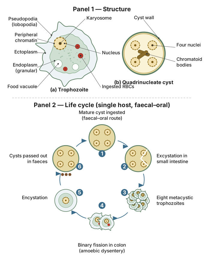
Figure — Two-panel labelled figure of Entamoeba histolytica. Panel 1 (structure) Entamoeba histolytica is a shapeless, pseudopodia-forming parasitic amoeba of the human intestine, with a direct (single-host) life cycle alternating between an active, disease-causing trophozoite stage and a resistant, transmissible cyst stage.
Structure of Entamoeba histolytica:
- Shape: No definite/fixed shape — it constantly changes shape by extending temporary, finger-like projections called pseudopodia ('false feet'), which are also used for locomotion and for engulfing food (phagocytosis).
- Size: Microscopic, typically 15-30 µm.
- Nucleus: A single, small, spherical nucleus with a centrally placed karyosome and a thin, evenly beaded peripheral chromatin layer (visible on staining).
- Cytoplasm: Differentiated into an outer clear ectoplasm (used to form pseudopodia) and an inner granular endoplasm (containing the nucleus, food vacuoles, and — in the pathogenic trophozoite stage — often engulfed host red blood cells).
- No mitochondria and no contractile vacuole — an adaptation to its low-oxygen intestinal habitat and its isotonic internal environment (living in a host, it does not need to expel excess water like free-living freshwater protozoans).
- No cell wall/pellicle, only a delicate plasma membrane, allowing shape change.
Life cycle (a direct, single-host cycle):
- Infection: A human ingests food or water contaminated with mature, resistant quadrinucleate cysts (cysts containing 4 nuclei) shed in the faeces of an infected person.
- Excystation: In the small intestine, the cyst wall is digested and the cyst releases a single amoeba that divides to form 8 small trophozoites (metacystic trophozoites).
- Trophozoite stage (active, pathogenic stage): These trophozoites migrate to and colonise the large intestine (colon), where they feed by phagocytosis, multiply by simple binary fission, and in pathogenic infections invade the intestinal mucosa, causing ulceration — this is the stage responsible for amoebic dysentery (bloody, mucus-containing diarrhoea) and, if it spreads via the bloodstream, amoebic liver abscess. …
- CBSE 2023Set ANNUAL8 marksQ.Explain the structure and life cycle of Entamoeba histolytica with the help of neat and labelled diagrams.
›Reveal solutionSolution
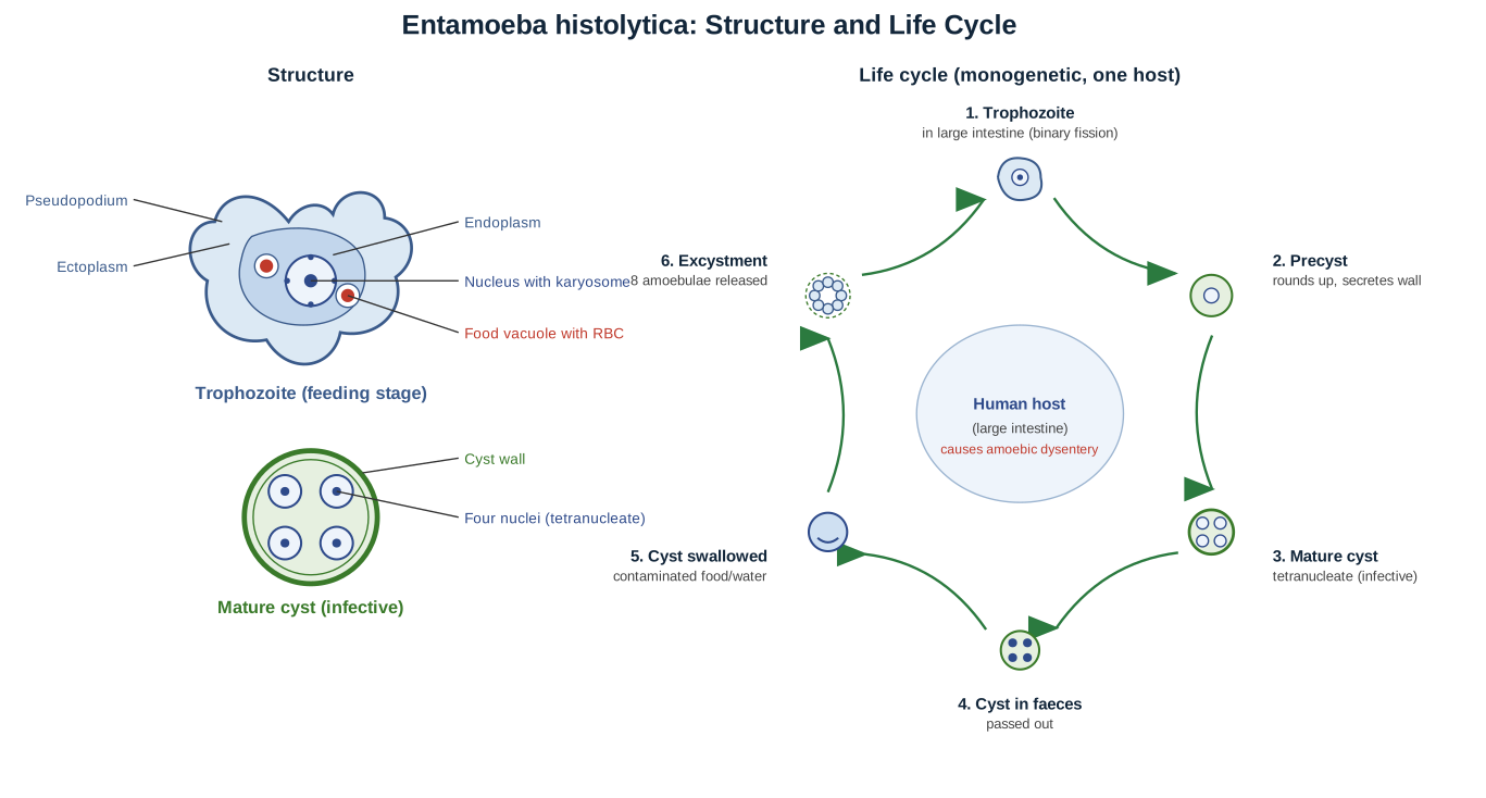
Entamoeba histolytica: trophozoite (ectoplasm, endoplasm, pseudopodium, single nucleus with central karyosome, food vacuoles with ingested RBCs) and mature tetranucleate cyst (cyst wall, four nuclei), with the monogenetic life cycle from trophozoite to precyst to cyst to excystment. Entamoeba histolytica is a monogenetic gut parasite of humans causing amoebic dysentery; it has a trophozoite (feeding) stage and a resistant cyst stage, and spreads by cysts through contaminated food and water.
(Diagrams to be drawn and labelled: (1) the TROPHOZOITE - showing ectoplasm, endoplasm, pseudopodia, a single nucleus with a central karyosome, and food vacuoles with ingested RBCs; (2) the mature CYST - a rounded, four-nucleate (tetranucleate) cyst with a cyst wall; and (3) the LIFE-CYCLE stages in sequence.)
Structure of Entamoeba histolytica:
- It is an endoparasitic protozoan living in the large intestine (colon) of humans.
- The trophozoite (active, feeding form) is amoeboid, with an outer clear ECTOPLASM and inner granular ENDOPLASM.
- It forms blunt PSEUDOPODIA for creeping movement and for engulfing food (including red blood cells and tissue).
- It has a single spherical NUCLEUS with a central karyosome (endosome) and peripheral chromatin.
- It has food vacuoles but NO contractile vacuole (being a parasite in an isotonic medium).
Life cycle (monogenetic - completed in a single host, man):
- Trophozoite stage:
-
The trophozoites live in the wall/lumen of the large intestine, feed on the mucosa and RBCs, cause ulcers, and multiply by binary fission. This produces amoebic dysentery (blood and mucus in stools).
- Encystment (cyst formation):
-
Some trophozoites become rounded, throw out food, and secrete a resistant CYST WALL around themselves, forming a precyst.
-
Within the cyst the nucleus divides twice to form a mature TETRANUCLEATE (four-nucleate) cyst - the infective stage.
- Transmission:
-
The mature cysts pass out with the FAECES of the patient.
-
Through contaminated food, water or hands (poor sanitation; house flies may act as carriers), the cysts are SWALLOWED by a healthy person.
- Excystment and multiplication: …
- CBSE 2019Set ANNUAL8 marksQ.Explain the structure and life-cycle of Entamoeba histolytica with the help of neat and labelled diagrams.
›Reveal solutionSolution
Figure — Two-panel labelled figure of Entamoeba histolytica. Panel 1 (structure) Entamoeba histolytica is an amoeboid gut parasite that alternates between a feeding/pathogenic trophozoite stage in the host's colon and a resistant, quadrinucleate cyst stage that is passed in faeces and transmits the infection through contaminated food or water.
Structure:
- Trophozoite (feeding stage): Unicellular, with no definite/fixed shape (amoeboid), moves and feeds using blunt, finger-like pseudopodia (lobopodia). Cytoplasm is differentiated into a clear outer ectoplasm and a granular inner endoplasm (which often contains ingested/phagocytosed red blood cells in the pathogenic form). It has a single, small nucleus with a characteristic centrally placed karyosome and peripheral chromatin granules. No cyst wall is present at this stage; it thrives in the moist environment of the large intestine.
- Cyst (resting/infective stage): Spherical, surrounded by a thick, resistant wall that protects it from the external environment and the host's gastric acid. Young cysts are uninucleate, maturing to the fully infective quadrinucleate (4-nucleate) cyst; may also contain sausage-shaped chromatoid bodies (stored glycogen/RNA-protein food reserves) — more prominent in immature cysts.
Life cycle:
- Infection: A human host ingests mature quadrinucleate cysts present in faecally contaminated food or water.
- Excystation: In the small intestine, the cyst wall is digested, releasing a single amoeba which undergoes nuclear and cytoplasmic division to give rise to 8 small metacystic trophozoites. …
🎓Unlock everything free for 14 days
- ✓Full step-by-step solutions
- ✓Concept-first explanations
- ✓Methods, shortcuts & mistakes
- ✓PYQ mapping + timed mock tests
Full access for 14 days. No credit card required.