Q.Explain internal structure of kidney with the help of suitable diagram.
You're viewing a preview — the full solution, concept, methods & PYQ mapping are locked.
Start your 14-day free trial to unlock the full solution →The kidney has an outer cortex (containing Malpighian bodies and convoluted tubules), an inner medulla (pyramids with loops of Henle and collecting ducts, opening at papillae into minor then major calyces), and a pelv...
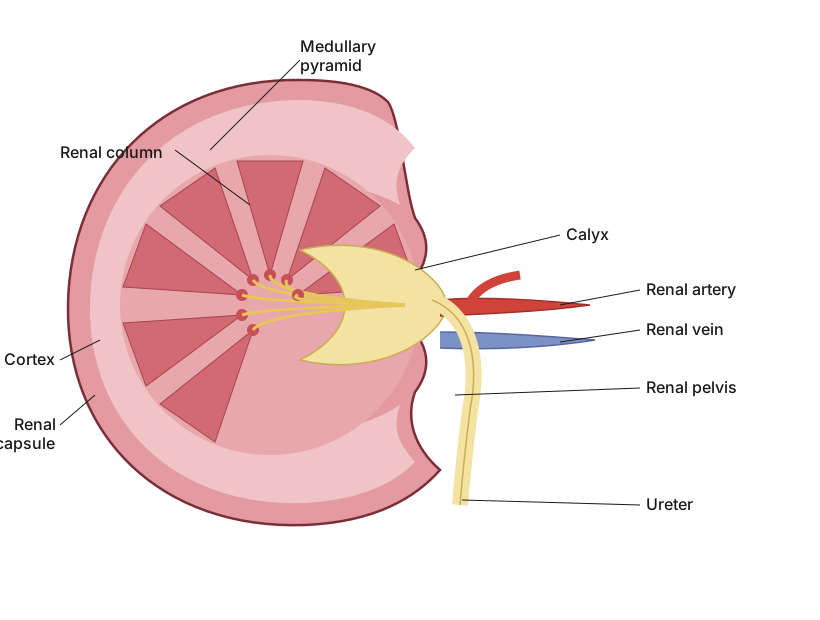
Kidney = outer cortex + inner medulla (pyramids) + pelvis draining into the ureter.
A longitudinal section through the human kidney reveals three principal regions. The outer, granular-looking Cortex lies just beneath the fibrous capsule and contains the Malpighian bodies (renal corpuscles — the glomerulus enclosed in Bowman's capsule) along with the proximal and distal convoluted tubules of each nephron. The inner Medulla appears striated and is organised into a number of cone-shaped Renal (Malpighian) pyramids, whose bases face the cortex and whose apices (papillae) point inward; the medulla contains the loops of Henle and the collecting ducts of the nephrons. Urine draining from the collecting ducts at the tip (papilla) of each pyramid empties into a cup-shaped Minor calyx; several minor calyces unite to form 2-3 larger Major calyces, which in turn open into a single funnel-shaped cavity, the Pelvis, at the centre of the kidney. The pelvis narrows and continues out of the kidney (at the hilum) as the Ureter, which carries urine down to the urinary bladder. Also at the hilum …
Unlock everything free for 14 days
- Full step-by-step solutions
- Concept-first explanations
- Methods, shortcuts & mistakes
- PYQ mapping + timed mock tests
Full access for 14 days. No credit card required.