Q.Sketch and label 'L.S. of human kidney'.
You're viewing a preview — the full solution, concept, methods & PYQ mapping are locked.
Start your 14-day free trial to unlock the full solution →The kidney in L.S.
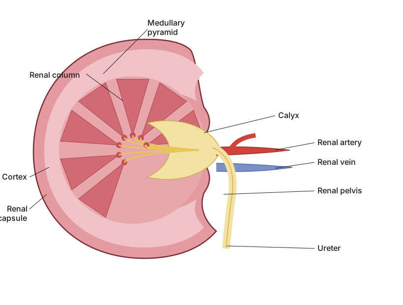
A longitudinal section of the kidney shows an outer cortex, inner medulla with pyramids, and a central pelvis draining into the ureter.
In longitudinal section, the human kidney is bean-shaped, with a convex outer border and a concave inner border bearing a notch called the hilum, through which the renal artery enters, and the renal vein and ureter exit. The kidney tissue is organised into two main zones: an outer, reddish-brown cortex, which contains the Malpighian corpuscles (glomerulus + Bowman's capsule) and the proximal and distal convoluted tubules; and an inner, paler medulla, which is arranged into a number of cone-shaped renal pyramids, each containing the loops of Henle and collecting ducts, with cortical tissue (columns of Bertini) dipping between adjacent pyramids. The apex (papilla) of each pyramid projects into and opens through several openings into a cup-like minor calyx; several minor calyces unite to form a major calyx, and the major calyces converge into a single funnel-shaped renal pelvis, which continues as …
Unlock everything free for 14 days
- Full step-by-step solutions
- Concept-first explanations
- Methods, shortcuts & mistakes
- PYQ mapping + timed mock tests
Full access for 14 days. No credit card required.