Q.Explain with the help of a suitable diagram conducting system of human heart.
OR
Give reasons :
(A) Lymphatic vessels are milky in appearance.
(B) Monocytes are called scavengers.
(C) The wall of left ventricle is thicker than right ventricle.
(D) Valves are present in the veins.
(E) Pulmonary veins carry oxygenated blood.
You're viewing a preview — the full solution, concept, methods & PYQ mapping are locked.
Start your 14-day free trial to unlock the full solution →The heartbeat is coordinated by nodal tissue: SA node (pacemaker, right atrium) -> AV node (base of right atrium) -> Bundle of His -> Purkinje fibres spreading through the ventricles, giving a coordinated, rhythmic co...
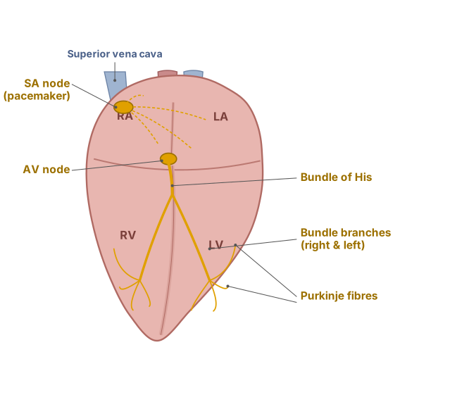
SA node -> AV node -> Bundle of His -> Purkinje fibres coordinates the heartbeat. OR: five reasoned facts about lymph, monocytes, ventricle walls, valves and pulmonary veins.
Conducting system of the human heart: The rhythmic heartbeat is initiated and coordinated by a specialised system of modified cardiac (nodal) tissue, not by nerves. (1) The Sino-atrial (SA) node, embedded in the wall of the right atrium near the opening of the superior vena cava, spontaneously and rhythmically generates electrical impulses at the fastest intrinsic rate of any cardiac tissue, so it sets the pace for the whole heart and is called the natural 'pacemaker'. (2) The impulse spreads through both atria, causing them to contract, and reaches the Atrio-ventricular (AV) node, located at the base of the right atrium near the interatrial septum; the AV node delays the impulse briefly, allowing the atria to finish emptying blood into the ventricles before ventricular contraction begins. (3) From the AV node, the impulse passes into the Bundle of His (atrioventricular bundle), a tract of specialised fibres that penetrates the fibrous septum separating atria from ventricles and then divides into right and left bundle branches running down either side of the interventricular septum. (4) These bundle branches give rise to fine, fast-conducting Purkinje fibres that spread throughout the walls of both ventricles, ensuring the impulse reaches all parts of the ventricular muscle nearly simultaneously, producing a strong, coordinated, single contraction of both ventricles together, rather than a disorganised twitch.
OR — Give reasons:
(A) Lymphatic vessels are milky in appearance because the lacteals (lymphatic vessels in the small intestine) absorb digested fats in the form of tiny fat globules (chylomicrons) from the gut; this fat-laden lymph, called chyle, has a characteristic milky-white colour.
(B) Monocytes are called 'scavengers' because they are strongly phagocytic white blood cells that engulf and digest bacteria, dead/damaged cells, and cellular debris; after migrating into tissues they mature into macrophages, which continue this clean-up/scavenging role throughout the body. …
Unlock everything free for 14 days
- Full step-by-step solutions
- Concept-first explanations
- Methods, shortcuts & mistakes
- PYQ mapping + timed mock tests
Full access for 14 days. No credit card required.